Imaging with IMAG’IC, the photonic imaging core-facility of Institut Cochin, Paris, France.
Posted by FocalPlane, on 20 February 2024
In our ‘Imaging with…’ blog post, we meet the staff of IMAG’IC, the photonic imaging core-facility of Institut Cochin, Paris, France.
https://institutcochin.fr/plateformes/imagic
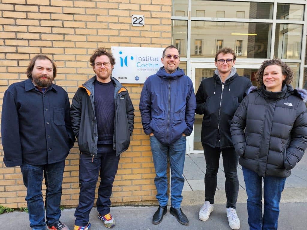
Staff role call (from left to right):
Gabriel Le Goff, Image processing, Engineer
Expertise: Finding or creating new algorithms to analyze users’ acquisitions.
Most likely to be found praying that my algorithms will work and swearing when they don’t.
Thomas Guilbert, PhD, Optical Developments, Research Engineer
Expertise: 2P microscopy, quality control, data handling, repairing with tape and twine ends
Most likely to be found aligning lasers while listening to (sometimes) strange music.
Pierre Bourdoncle, PhD, Core Facility Manager, Research Engineer
Expertise: Solving human, administrative, microscope and IT issues… while trying to anticipate future ones!
Most likely to be found yelling at the machines that don’t work, when I’m not managing our great team.
Raphaël Braud-Mussi, Image Processing, Engineering Informatics
Expertise: Pre and post process algorithm to exploit machine learning data in a good way.
Most likely to be found coding, making some *bee boop* noises.
Julie Lesieur, Biology Engineering
Expertise: cell culture, tissue and sample preparation
Most likely to be found telling users to stop using mounting media containing DAPI!
Microscope role call:
Leica SP8 DIVE FLIM BDSL2, Leica SP5 2P, Leica SP8X STED FLIM BDSL2, Leica GSD, Spinning-Disk X1 (SD) (x3 – BDSL1, 2 and 3), SD Evident W1, Till Photonic iMIC, Leica DMI6000, Zeiss Observer 7 Inscoper driven, Zeiss Axiovert 200M, Olympus BX63, ImageStreamX MKII, Nikon AZ100
Who can access the facility?
We are an open-access collaborative facility enabling researchers from within Institut Cochin and elsewhere to perform cutting-edge studies using state-of-the-art equipment. This includes research on diverse samples like cells or tissues, both in vitro and in vivo. Many of the instruments are located in biosafety levels 2 and 3 laboratories, which allows imaging of live cells, tissue samples of human origin and/or infected with pathogens, and whole mammals infected with communicable diseases.
All our systems are accessible via a CID image database for backup, archiving, processing, analysis & sharing.
We are very attached to our different networks which are an inexhaustible source of useful knowledge for our businesses, locally (RTmfm, GDR Imabio -MiFoBio & France-BioImaging) and internationally (QUAREP-LiMi & EuroBioImaging).
Pet peeve (something that users do that is annoying): We still don’t understand how pools of immersion oil end up on dry objectives. The plot thickens…
Favourite microscope: We’re still looking for the ideal microscope capable of giving us its health status over time to improve reproducibility in research 😉
Favourite thing to image: Anything that is fluorescent under the right excitation, from candies to kinin, with or without gin!
Best bit of advice (that you give or have been given): If it won’t hold with gaffer tape, it will with more gaffer tape.
If money was no object, we would buy… Definitely more space…
Can you give us some examples of recent papers that were published with your assistance?
- Nicolas-Boluda, A., Vaquero, J., Vimeux, L., Guilbert, T., Barrin, S., Kantari-Mimoun, C., Ponzo, M., Renault, G., Deptula, P., Pogoda, K., Bucki, R., Cascone, I., Courty, J., Fouassier, L., Gazeau, F., & Donnadieu, E. (2021). Tumor stiffening reversion through collagen crosslinking inhibition improves T cell migration and anti-PD-1 treatment. eLife, 10, e58688. https://doi.org/10.7554/eLife.58688
- Faklaris, O., Bancel-Vallée, L., Dauphin, A., Monterroso, B., Frère, P., Geny, D., Manoliu, T., de Rossi, S., Cordelières, F. P., Schapman, D., Nitschke, R., Cau, J., & Guilbert, T. (2022). Quality assessment in light microscopy for routine use through simple tools and robust metrics. Journal of Cell Biology, 221 (11), e202107093. https://doi.org/10.1083/jcb.202107093
How should users acknowledge the facility and why is it important?
Within Institut Cochin, each user signs a charter common to all our 10 core facilities, which requires that any significant contribution to a research project is endorsed by an article author signature. Acknowledgement is also required as soon as a machine in IMAG’IC has been used. This is very important for us in order to gain visibility within the scientific community, for our National Infrastructure in Biology and Health (France BioImaging) and also for our potential funders.
 (No Ratings Yet)
Get involved
Create an account or log in to post your story on FocalPlane.
More posts like this

Filter by
- NewsApply
- DiscussionsApply
- How toApply
- ToolsApply
- Case studiesApply
- InterviewsApply
- JobsApply
- EducationApply
- Blog seriesApply
- Latin American Micro..scopistsApply
- Bio-image Analysis w..ith NapariApply
- Imaging with…Apply
- Towards Global Acces..sApply
- Latin America Bioima..gingApply
- From Zero to Qupath ..HeroApply
- Asian Microscopists ..and Cell BiologistsApply
- AIC at HHMI JaneliaApply
- Deep Learning for Bi..o-image analysisApply
- GloBIAS – updates fr..om the communityApply
- WAMBIAN: West Africa.. in FocusApply
- Volume EMApply
- Highlights from Euro..-BioImagingApply
- LSFM seriesApply
- DIY MicroscopyApply
- View all