FocalPlane/elmi2024 image competition – vote for your favourite
Posted by FocalPlane, on 3 June 2024
Thanks to everyone who entered our FocalPlane/elmi2024 image competition, we were delighted to receive so many wonderful entries. The organisers of elmi2024 and the JCS team have shortlisted 10 images for our public vote. Thanks to Liam Rooney for coordinating the voting from the elmi2024 team, as well as the rest of the organising committee, Kurt Anderson, Siân Culley, Joëlle Goulding, Marco Marcello, Gail McConnell, Peter O’Toole, Tim Self, Jessica Valli and Kate Wooding.
You can vote using the poll at the bottom of this page. The image with the most votes will be featured on the cover of Journal of Cell Science and the scientist will win £150. Voting will be open from now until the end of elmi2024 (Friday 7 June), but you don’t need to be attending in order to vote.
Voting has now closed.
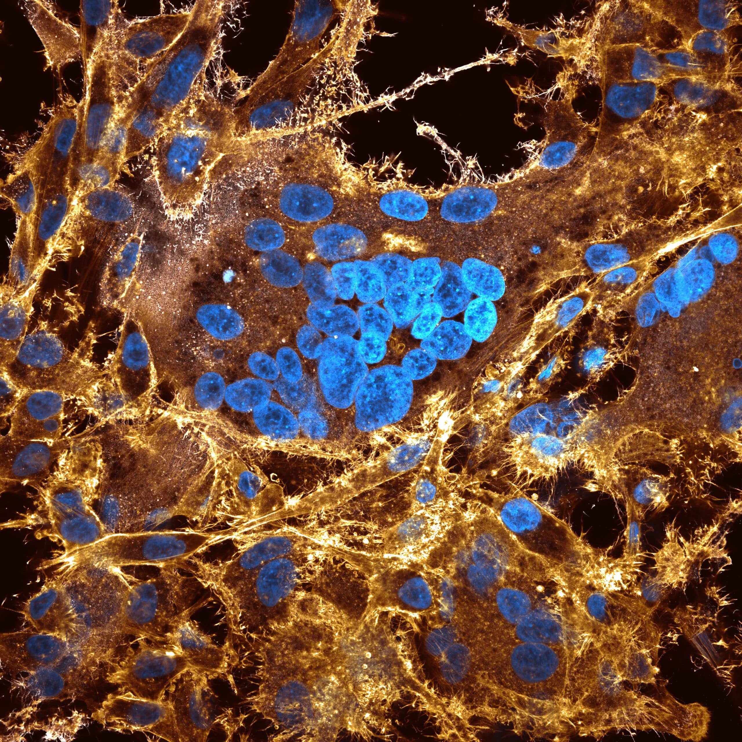
1. Boa constrictor kidney syncytium – Joe McKellar
This image is that of a cell culture of boa constrictor kidney cells that are overexpressing a viral fusogenic glycoprotein (VSV-G), which causes cells to fuse together, forming a syncytium. It was acquired using an LSM 980 8Y confocal with an Airyscan 2 module. The image has been pre-processed in the Zen Blue software with post-processing using FIJI. F-actin = gold; cellular DNA = cyan and VSV-G = white.
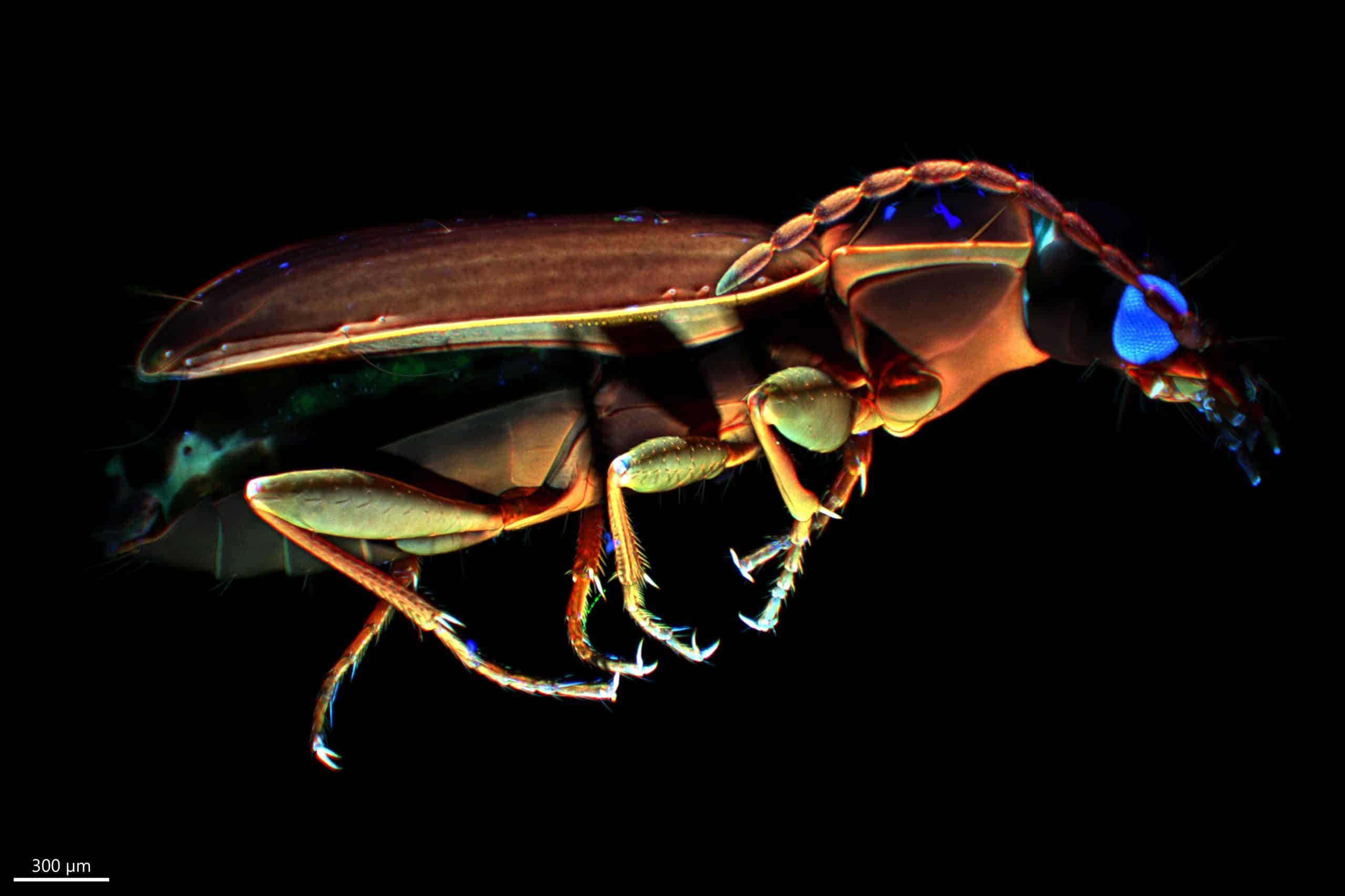
2. Beetle – Jiří Černý
Thanks to the natural autofluorescence of chitin, the main component of the cuticle of insects, these arthropods can be displayed in a different form under fluorescence microscopes. Imaged using light-sheet fluorescence microscopy and Imaris and HuygensPro softwares for postprocessing and reconstruction.
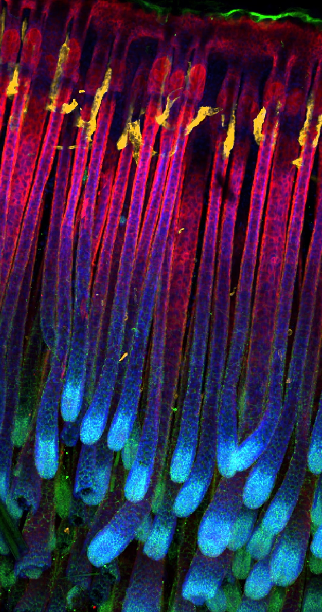
3. Mouse hair follicles in cleared postnatal skin tissue – Marina Schernthanner
3D whole-mount immunofluorescence image of cleared murine skin tissue (postnatal day 9), side view. Keratin 14 (basal epidermal cells) in red, P-Cadherin (keratinocytes) in green, LYVE-1 (lymphatics) in yellow and DAPI (nuclei) in blue. The image was acquired using an Andor Dragonfly spinning disk microscope and processed in Imaris 9.5.1.

4. Anarchy in the cell – Alan Marron
An image of hornwort (Anthoceros agrestis) cells showing chlorophyll autofluorescence (chloroplasts) in magenta and mVenus-tagged FtsZ protein in yellow. This shows FtsZ filaments forming across the single chloroplast of hornwort cells.
This image was taken on a Leica SP8 upright confocal microscope. A maximum intensity projection of the z-stack was produced in LasX and the brightness and contrast adjusted using Fiji/ImageJ.
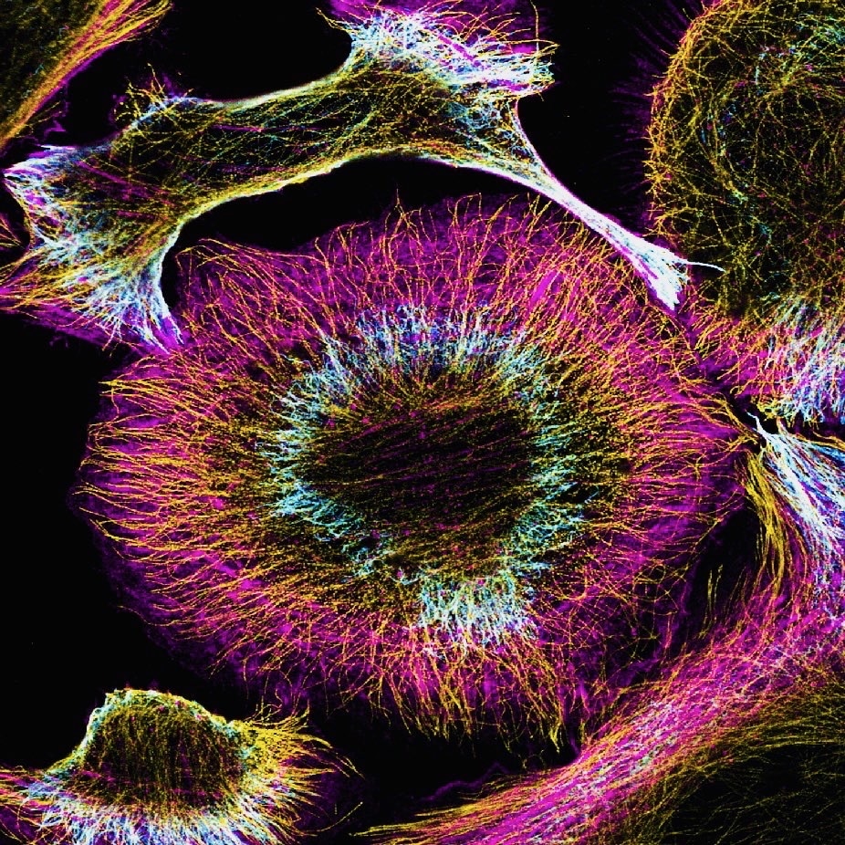
5. The skeleton – Lorna Young
U2OS cell stained with actin (magenta), microtubules (yellow) and vimentin (intermediate filaments). Imaged on Zeiss 900 Airyscan.
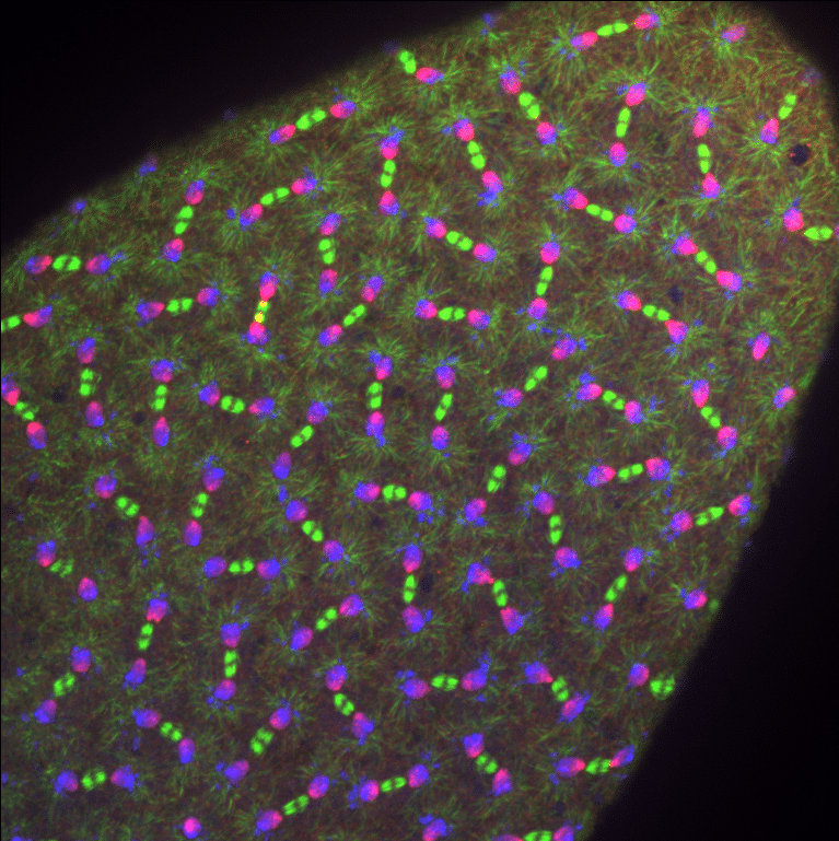
6. Synchronised division – Jovan Brockett
The image is of a Drosophila melanogaster embryo undergoing syncytial nuclear divisions. Thus, all of the nuclei are in sync with each other and, in this image, are in anaphase. The labels are alpha-tubulin in green to mark the microtubules and mitotic spindles. The red is phosphorylated histone (pH3). The blue is a nuclear marker called DAPI.
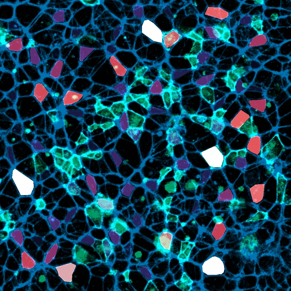
7. Mosaic – Ioakeim (Makis) Ampartzidis
In this image, the actin cytoskeleton of neuroepithelial cells is shown in blue, and the membranes of some randomly selected cells are highlighted in cyan using synthetic fluorescent labelling dyes. To emphasize the cellular variability within our neuroepithelial sheets, the apical membranes of cells were pseudo-coloured based on their size, ranging from purple (small apical area) to white (large).
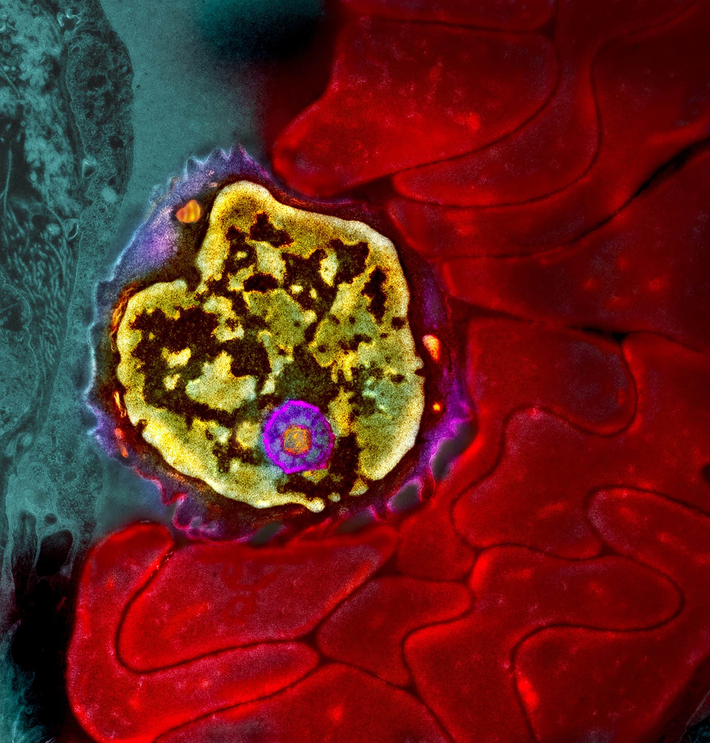
8. Glorious lymphocyte – Rossana Melo
A leukocyte (lymphocyte) trapped among aggregates of red blood cells in a small lung vessel of a patient with asthma.
The image was acquired with a transmission electron microscope (Tecnai G2 Spirit Bio Twin ThermoFischer Scientific) at 80 KV and colorized using Photoshop.
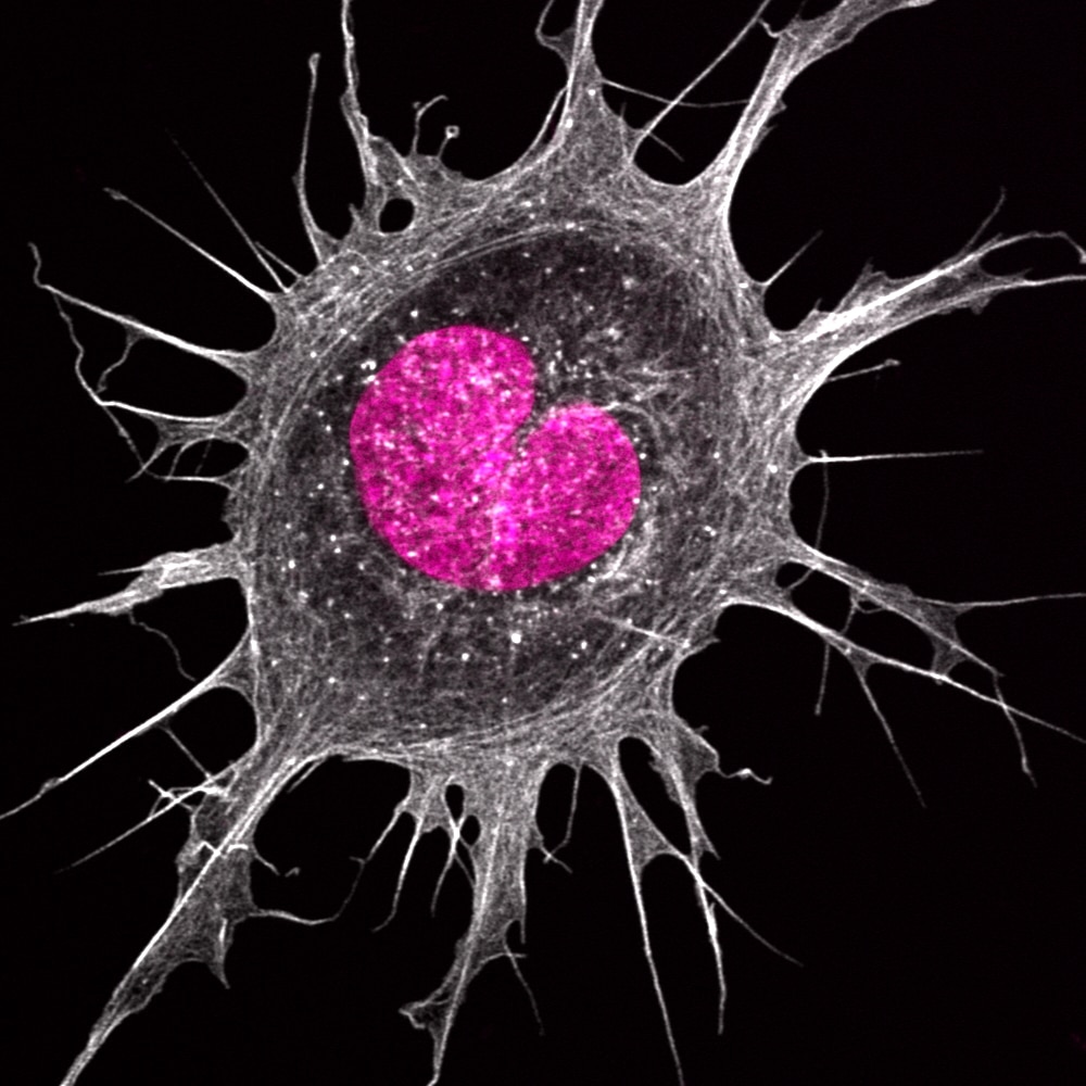
9. At the heart of a cancer cell – Hoang Anh Le
An Ewing’s sarcoma cancer cell with a curious heart-shaped nucleus. Imaged using Zeiss880 with Airyscan.
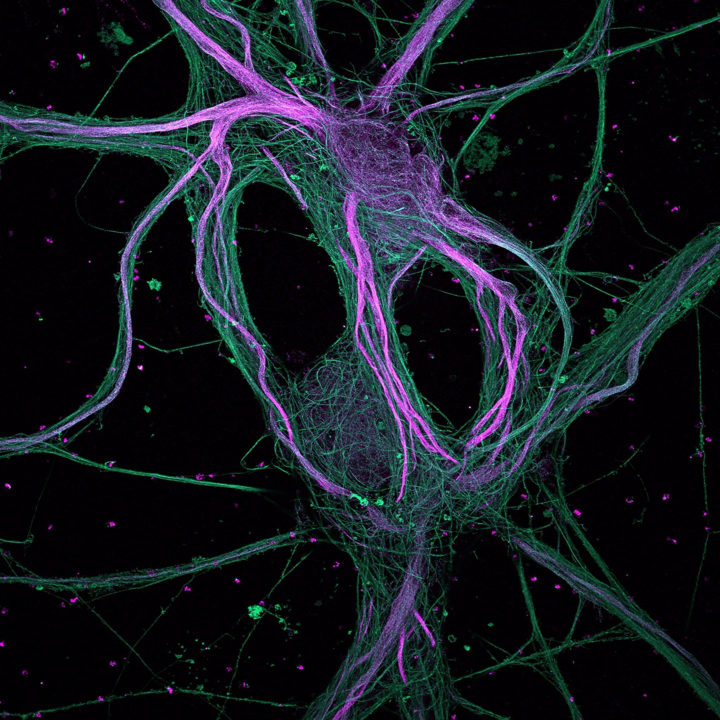
10. You are always on my mind – Ciarán Butler-Hallisey
4x Expanded (U-ExM) rat hippocampal neurons showing microtubules in green and Map2 in magenta. Imaged using spinning disk confocal microscopy.
Thank you for voting
 (48 votes, average: 1.00 out of 1)
44 thoughts on “FocalPlane/elmi2024 image competition – vote for your favourite”
Leave a Reply
Get involved
Create an account or log in to post your story on FocalPlane.
More posts like this

Filter by
- NewsApply
- DiscussionsApply
- How toApply
- ToolsApply
- Case studiesApply
- InterviewsApply
- JobsApply
- EducationApply
- Blog seriesApply
- Imaging with…Apply
- Towards Global Acces..sApply
- Latin America Bioima..gingApply
- From Zero to Qupath ..HeroApply
- Asian Microscopists ..and Cell BiologistsApply
- AIC at HHMI JaneliaApply
- Deep Learning for Bi..o-image analysisApply
- GloBIAS – updates fr..om the communityApply
- WAMBIAN: West Africa.. in FocusApply
- Volume EMApply
- Latin American Micro..scopistsApply
- Bio-image Analysis w..ith NapariApply
- Highlights from Euro..-BioImagingApply
- LSFM seriesApply
- DIY MicroscopyApply
- View all
Perfect!
Wonderful
Science and art in one image
Incrível
Indescritível
Parabéns pelo excelente trabalho. Orgulho!
Impacting image
Electron microscopy at its best.
Number 8
Seria ideal que cada imagem trouxesse informações sobre importância para a Ciência.
Como
Perfeito. Muito conhecimento transmitido.
Número 8 é visivelmente real .
Perfect image!
Número 8
A número 8 ficou sensacional!
Número 8
Gostei imagem de alta resolução.Parabens número 8
Sensacional número 8
Número 8 é maravilhoso
Excelente trabalho parabéns
Parabéns Rossana!
Maravilhosa photo 8
O número 8 é fantástico!
Wonderful
Número 8
8
8
Número 8
Número 8
Loved number 8
Número 8
Foto 8.
Perfect
Número 8
8
Number 8
Numero 8. Linda foto. Parabéns.
Numero8. Linda foto.
Número 8 está sensacional!
8. Glorious lymphocyte – Rossana Melo
My choice is number 8 but all of them are unique and very pleasant to look.
All very beautiful but the number 8 is stunning!
2
A número 8 ficou sensacional.
Imagens instigantes. A tecnologia leva-nos ao inimaginável.