Interview with Dr Benjamin Compans: From Academia to Industry in Super-Resolution Microscopy
Posted by Sally Horton, on 7 May 2025

In this interview, I sat down with Dr Benjamin Compans, A field Application Scientist at Abbelight (pictured above).
Microscopy is more than just a tool; it’s a window into the intricate and often unseen world of biological structures. For many researchers, their first encounter with a microscope can be a defining moment in their scientific journey. In this interview, I sit down with Dr Benjamin Compans, a former academic researcher who played a pivotal role in my own introduction to microscopy.
When we first met, Ben was deep into his postdoc within the Burrone lab, at the Centre for Developmental Neurobiology, King’s College London. Here, Ben was using advanced imaging techniques to push the boundaries of our understanding of synapses and the organisation of neurotransmitter receptors in neurons. Ben’s expertise and passion for microscopy was infectious, and I was lucky to learn from him early in my own career. Not only did Ben teach me about Stochastic Optical Reconstruction Microscopy (STORM), a single molecule localisation microscopy (SMLM) technique, but he also greatly enhanced my overall understanding of the imaging process, helping me appreciate the nuances of each step, from sample preparation to data analysis. I applied this knowledge throughout my PhD, and now in my current postdoc at the Francis Crick Institute, where I continue to work with various imaging techniques.
Today, Ben has transitioned from academia into the industry, working at Abbelight, a microscopy company specialized in super-resolution microscopy.
In this conversation, we discuss Ben’s journey from the research lab to the corporate world, the evolution of microscopy, and their insights on where the field is headed. We also explore the lessons he has learned along the way and his advice for researchers looking to incorporate cutting-edge imaging techniques into their work.
Interview:
Thank you, Ben, for agreeing to take part in this interview, I’m excited to learn more about your experiences and insights in the field of microscopy. Can you start by telling us about your first experience with microscopy?
My first experience with microscopy took place during a short internship in the middle of my master’s degree, when I visited Dr. Daniel Choquet’s lab at the Interdisciplinary Institute for Neuroscience (IINS) in Bordeaux. There, I had the opportunity to meet Dr. Eric Hosy and his student, who were performing Single Particle Tracking (SPT) using a custom-built microscope. I vividly remember seeing small, fluorescent dots, each representing an individual AMPA receptor, moving along neuronal dendrites. For those unfamiliar with neuroscience, AMPA receptors are a type of glutamate receptor responsible for fast excitatory synaptic transmission. Two things fascinated me during that experience. First, I was amazed to see how these receptors diffused randomly via Brownian motion, yet occasionally stopping completely at synapses, where they would carry out their role in synaptic transmission. Second, I was deeply impressed by the ingenious methods researchers had developed to overcome the diffraction limit, a barrier in conventional fluorescence microscopy that would otherwise prevent us from visualizing individual molecules and gaining such detailed insights. This experience immediately inspired me to join this lab for my PhD.
When we first met, you were doing a postdoc. What was your research focus at the time, and how did microscopy play a role in it?
As you know, I was conducting my postdoctoral research in the lab of Prof. Juan Burrone at King’s College London. My work focused on understanding the nanoscale organization of a unique type of inhibitory synapse located along the axon initial segment of neurons. To give some context: back in 2013, three laboratories, including the one where I completed my PhD, used super-resolution microscopy to reveal that synaptic proteins at excitatory synapses are not randomly distributed, but instead clustered into nanodomains smaller than 100nm1–3. Similar findings were made at inhibitory synapses4. These discoveries significantly advanced our understanding of the molecular mechanisms that fine-tune synaptic transmission. What’s important to note is that most of these studies were conducted using in vitro models. While these models are essential for gaining fundamental insights, they do not capture the full complexity and elegance of synaptic connectivity and diversity found in the brain. My research aimed to apply the super-resolution imaging techniques I learned during my PhD to study a specific synapse type, which cannot be effectively studied in vitro, but are believed to play a critical role in neuronal circuit function.
Who or what had the biggest influence on your microscopy journey during your academic career?
My PhD supervisor, Dr. Eric Hosy, taught me a lot about advanced microscopy techniques such as Single Particle Tracking and SMLM. But more importantly, he taught me that despite the excitement of developing new methods, the importance is the biological question being asked. There are often multiple techniques that can be used together to address a single question, and if the right technique doesn’t yet exist, then it means that there is an opportunity for a new technical breakthrough
You work with super-resolution microscopy. What excites you the most about this technology?
Of course, super-resolution microscopy enables the creation of stunning images, but as a researcher, what excites me most about this technique is the level of precision it offers compared to more conventional methods. The gain of precision when measuring structure size, number or finally seeing the real organization of your favourite protein within a compartment. ‘Seeing is believing’, and the more detail you can extract from an image, the more accurately and confidently you can answer a scientific question.
What are some of the most exciting breakthroughs in microscopy that you’ve seen in recent years?
As a neuroscientist, two major examples immediately come to mind. First is the discovery of the nanoscale organization of synaptic proteins on both sides of the synaptic cleft, which plays a crucial role in fine-tuning synaptic transmission1–3,5. Second is the remarkable periodic organization of the cytoskeleton along neuronal axons6,7. Other fields of research have also greatly benefited from super-resolution microscopy, particularly in uncovering and tracking the precise organization of proteins and multiprotein complexes within various cellular compartments and organelles8.
What challenges still need to be addressed in super-resolution imaging?
A great deal of technical progress has been made over the past decade, reaching to Ångström resolution with fluorescence super-resolution imaging9, and I believe the next key step is to democratise these advanced imaging approaches for biologists. Researchers need to generate data across numerous targets and varied biological conditions, which demands more accessible, user-friendly, and automated tools, spanning everything from sample preparation to analysis software. While these techniques can achieve amazing resolution, when it comes down to biological sample, it is not always straightforward as a lot of factors will affect the final resolution, leading to artefacts if not well considered and the researcher to inexact conclusions. These risk that exist with any technique, and I strongly believe our role, working in industries developing such approach for biologist, is to make it the most straightforward and user-friendly to limit these risks.
How does working in a microscopy company differ from working in a research lab?
It really depends on the specific role within a microscopy company, as responsibilities vary greatly between teams like R&D and Customer Care. In my case, as a Field Application Scientist, my main role is to support researchers from diverse scientific fields. This involves understanding their research goals, proposing imaging solutions that could advance their work, and helping them use those solutions effectively to answer their biological questions. Unlike when I was in a research lab, I’m no longer the one conducting experiments, publishing papers, or applying for grants. However, I like to think that I can play an important role in supporting scientists through all of those steps by helping them to access advanced imaging techniques that will help them in their research. It’s an exciting and fulfilling mission to be part of a company that pays attention to the success of the researchers using our technology.
What advice would you give to PhD students or postdocs who want to incorporate advanced microscopy techniques into their research?
Like any technique, super-resolution microscopy requires some initial learning to understand how it works and how to use it effectively, from optimizing sample preparation to mastering available analysis tools. However, these techniques have evolved rapidly over the past decade, and now offer increasingly user-friendly solutions, thanks in large part to the work of several microscopy companies, including Abbelight. Support from these companies, especially through Field Application Scientists, can also be incredibly valuable for early-career researchers, providing guidance from experts who know how to apply these methods to real biological questions.
What motivated you to move from academic research into the microscopy industry?
Of course, I have to mention the difficulty young researchers face in securing permanent positions. However, in my case, I decided to join Abbelight after two years of collaborating with them. As a user of the Abbelight SAFe microscope during my postdoc at King’s College London, which you also used during your PhD, I had many discussions with the team at Abbelight. Through those interactions, I came to understand their mission and realized this could be a fantastic opportunity to contribute to the advancement of research by educating others about super-resolution microscopy using the expertise acquired during my PhD and postdoc, and helping them implement these techniques in their daily research. On top of that, working for an innovative French company founded by academic researchers is incredibly motivating, as they truly understand the challenges faced in research labs.
How did your PhD and postdoc experience prepare you for your current role?
My PhD and my postdoc allowed me to gain expertise on various super-resolution microscopy techniques. More importantly, they taught me how to identify when these approaches are best suited to address specific biological questions, and what advantages they offer over more conventional methods. During my academic career, giving talks, teaching courses, and mentoring students (like you), allowed me to be efficient at explaining both the technical and scientific aspect of microscopy. Additionally, I learned to manage multiple projects simultaneously, a skill that has proven essential in my current role.
What were some of the biggest challenges in transitioning from academia to industry?
As a PhD student or postdoc, you typically work independently (supervised, of course), but usually focused on your own project. At Abbelight, however, I interact with colleagues from various departments, including optical R&D, software development, and sales. This requires significant effort in communication, as we all come from different educational backgrounds and speak different ‘languages’. Although we belong to different teams, effective communication is essential for efficiency, from developing new products to helping scientists apply them in their research. It took me couple months to fully grasp and improve these communication skills, but I’ve really enjoyed the process. It has also given me the opportunity to learn from my colleagues.
Do you have a ‘favourite’ microscopy technique?
Stochastic Optical Reconstruction Microscopy (STORM) is still my favourite method. In my opinion, it is relatively easy to implement using standard fluorophores commonly found in the lab, making it an excellent entry point into super-resolution imaging techniques, whether for cell culture or thin tissue slices.
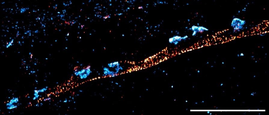
Do you have a ‘favourite’ image that you have acquired?
Yes, I still really like one image I captured during my postdoc at King’s College London using the Abbelight SAFe super-resolution system. It beautifully illustrates the nanoscale organisation of inhibitory synapses within the periodic structure of the Axon Initial Segment.
The image in question:
References:
- Nair, D. et al. Super-resolution imaging reveals that AMPA receptors inside synapses are dynamically organized in nanodomains regulated by PSD95. Journal of Neuroscience 33, (2013).
- MacGillavry, H. D., Song, Y., Raghavachari, S. & Blanpied, T. A. Nanoscale scaffolding domains within the postsynaptic density concentrate synaptic ampa receptors. Neuron 78, (2013).
- Fukata, Y. et al. Local palmitoylation cycles define activity-regulated postsynaptic subdomains. Journal of Cell Biology 202, (2013).
- Specht, C. G. et al. Quantitative Nanoscopy of Inhibitory Synapses: Counting Gephyrin Molecules and Receptor Binding Sites. Neuron 79, 308–321 (2013).
- Tang, A. H. et al. A trans-synaptic nanocolumn aligns neurotransmitter release to receptors. Nature 536, (2016).
- Leterrier, C. et al. Nanoscale Architecture of the Axon Initial Segment Reveals an Organized and Robust Scaffold. Cell Rep 13, (2015).
- Xu, K., Zhong, G. & Zhuang, X. Actin, spectrin, and associated proteins form a periodic cytoskeletal structure in axons. Science (1979) 339, (2013).
- Liu, S., Hoess, P. & Ries, J. Super-Resolution Microscopy for Structural Cell Biology. Annual Review of Biophysics vol. 51 Preprint at https://doi.org/10.1146/annurev-biophys-102521-112912 (2022).
- Reinhardt, S. C. M. et al. Ångström-resolution fluorescence microscopy. Nature 617, (2023).
 (No Ratings Yet)
Get involved
Create an account or log in to post your story on FocalPlane.
More posts like this

Filter by
- NewsApply
- DiscussionsApply
- How toApply
- ToolsApply
- Case studiesApply
- InterviewsApply
- JobsApply
- EducationApply
- Blog seriesApply
- Deep Learning for Bi..o-image analysisApply
- GloBIAS – updates fr..om the communityApply
- Volume EMApply
- Latin American Micro..scopistsApply
- Bio-image Analysis w..ith NapariApply
- Imaging with…Apply
- Towards Global Acces..sApply
- Latin America Bioima..gingApply
- From Zero to Qupath ..HeroApply
- Asian Microscopists ..and Cell BiologistsApply
- AIC at HHMI JaneliaApply
- Highlights from Euro..-BioImagingApply
- LSFM seriesApply
- DIY MicroscopyApply
- View all