Exploring the Microcosmos through Foldscope-Frugal Science Microscopy in Focus
Posted by Aarushi Verma, on 18 July 2025
Article from Aarushi Verma, Rubai Mushtaq, Kaavya K B, Imroz Alam, Neha Gajbhiye and Anupma Harshal W.
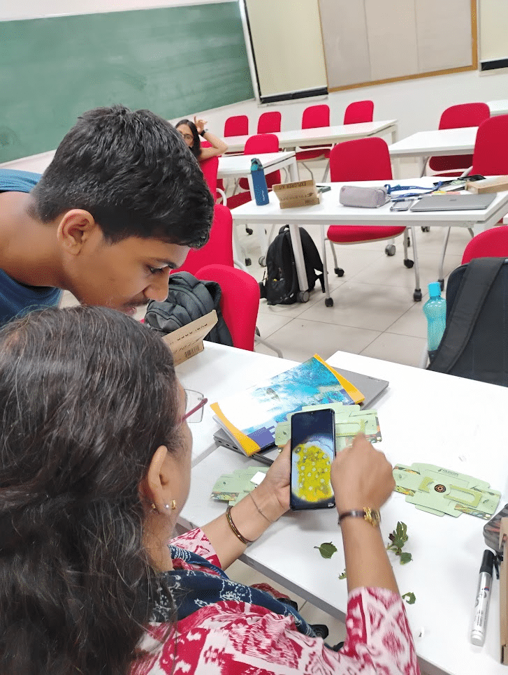
The third annual iteration of the Lodha Genius Programme at Ashoka University, Sonepat, featured a month-long workshop series for students of grades 9 and 10, introducing them to the intricate diversity of the microscopic world through the use of a frugal science innovation—the Foldscope. Much like the initiative described in this article on IndiaBioscience, this programme aimed to democratise science education by using frugal innovations to bridge access and ignite curiosity. The Foldscope’s lightweight, origami-inspired paper microscope, created by Manu Prakash, immediately captivated the students, sparking both scientific inquiry and creative reflection.
The workshop, conducted in rotating batches, was thoughtfully designed to introduce the next generation of scientists to the workings of the Foldscope while integrating foundational scientific concepts. These included antimicrobial resistance, visual classification and comparison of different vegetable cells, and the structural diversity of pollen grains from various flowers. Underlying themes such as the democratisation of scientific knowledge through frugal innovation and the ethics of scientific inquiry were interwoven throughout the sessions, encouraging critical reflection alongside hands-on exploration.
Each day began with a light-hearted “chirpy bird” dance, re-energising students after lunch — a ritual that foreshadowed the classroom’s ambience: rooted in movement, engagement, and hands-on learning. The first day introduced the concept of antimicrobial resistance through the metaphor of ‘superbugs,’ prompting students to reflect on its rise as a rising public health concern and its implications on low-resource societies. This session also emphasised the importance of civic responsibility in healthcare, cautioning students against the careless overuse of antibiotics and highlighting the critical role of completing prescribed antibiotic courses in addressing this global health challenge.
Frugality through and through!
The Foldscope exploration began with the observation of a premade fern rhizome slide included in each student’s kit. The classroom instantly filled with exclamations of wonder—“wows” and “aahs”—as students caught their first glimpse of the microscopic world. Staying true to the spirit of frugal science, even the sample preparation materials reflected innovation on a budget: sticky transparent tape replaced traditional cover slips, glass slides were washed and reused, and tweezers were replaced by fingernails and fingertips for peeling thin sample films. Students were also surprised to learn that stains weren’t necessary to visualise contrast — ultra-thin specimens and a strong light source worked as well.
Many students pointed out the potential concern that the transparent tape might interfere with the clarity. The first live biological sample—onion epidermal cells—was intentionally chosen to address this. The clear, brick wall-like structure of onion cells helped students easily distinguish between the cells and artefacts like air bubbles introduced by the tape or water early on. On day 2 they observed potato cells. Locating the starch-rich cells tested their fine adjustment skills as they worked towards getting a picture-worthy field of view.
Practising science communication through creative expression
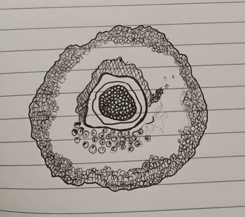
Each observation followed a standard protocol: beginning at 50x magnification, then progressing to 140x and 340x. Each magnification progressively revealed the underlying order and granularity of the sample. As part of good scientific practice, students documented their findings through both sketches and photographs. For some, drawing became a form of creative expression, while for others it was a tool to better reflect on structural features.
In addition, students shared their insights through personal blog posts on the official Foldscopes Microcosmos site, showcasing their work to the global Foldscopers community. All of these mediums gave the students a chance to express their voices and experiment with diverse forms of scientific communication.
Learning that comes alive!
Rooted in experiential learning, the workshop kept students actively engaged during the three-hour daily sessions. They relied entirely on manual techniques to create slides without conventional lab equipment. Despite the frugal setup, safety and hygiene protocols were strictly enforced — biological samples were restricted, and students cleaned their workspaces and washed their hands after every session.
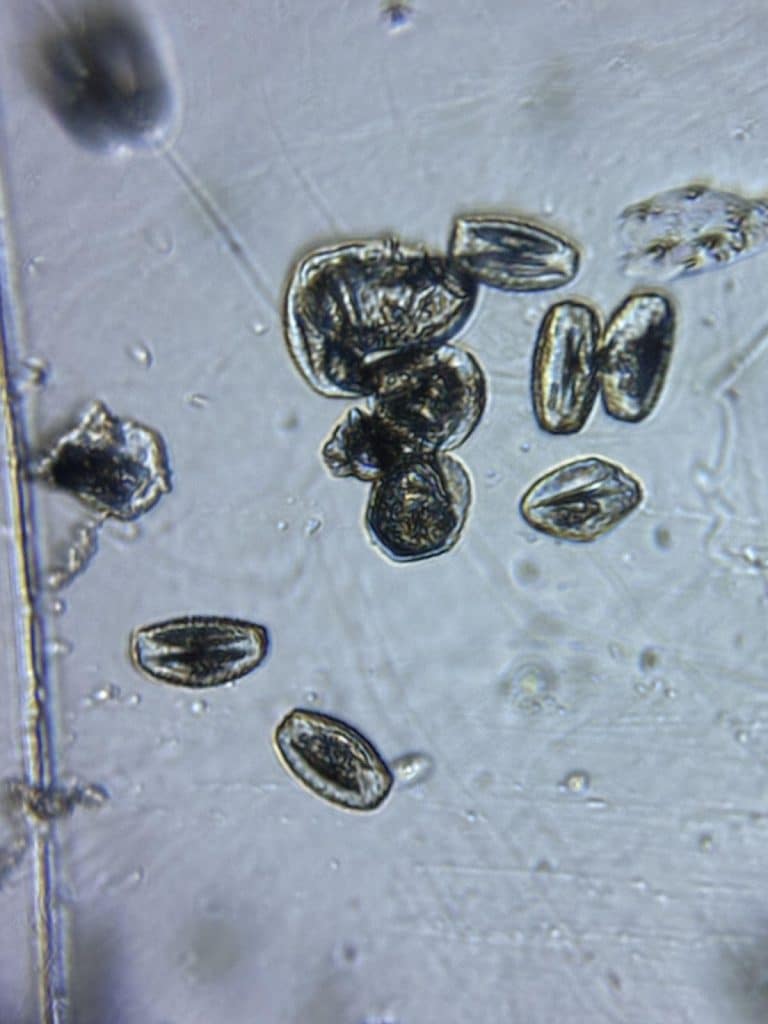
Day 3 was a busy one, as students explored pollen from flowers collected across campus — Magnolia, Bougainvillea, and Golden Shower. Observing differences in size, shape, and surface structure debunked the myth that all pollen are spherical, yellow, and smooth. This session also sparked discussions about pollinators based on pollen visibility and floral structure.
Students were introduced to the concept of magnification and how to report findings appropriately. Specimen observation was often a painstaking process; students discovered that the clearest observations came from isolating a single pollen grain within the field of view and developed patience, precision, and a sharper eye for microscopic complexity along the way.
Despite the intensity, students showed no signs of fatigue. Many were eager to continue beyond the scheduled class time, even redoing slide preparations to improve results. Their enthusiasm made it clear how rewarding this hands-on learning journey had been.
Students taking reins of their learning experience
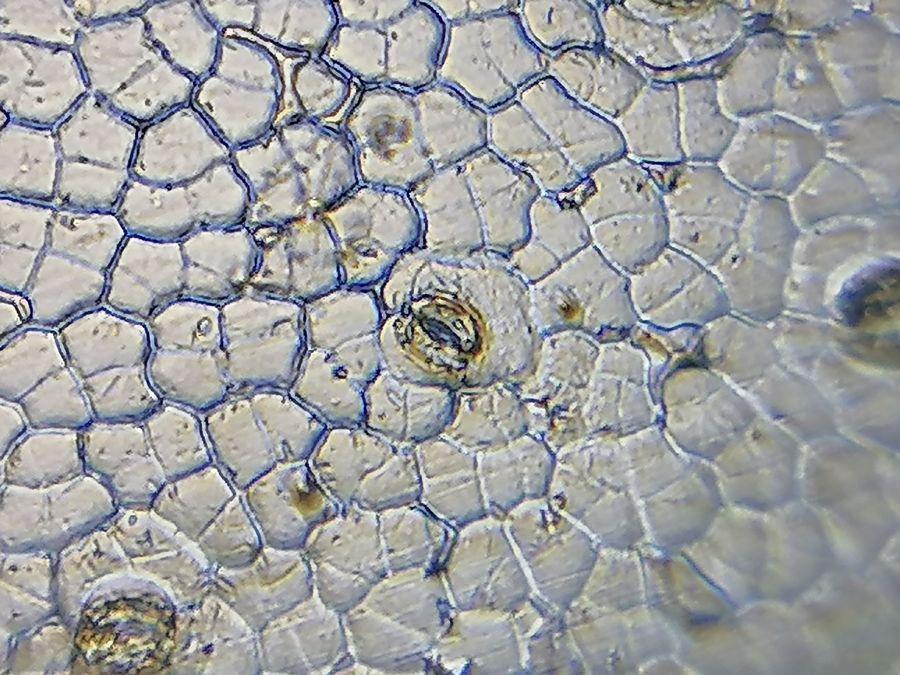
By day 4, students were adept at recognising that different specimens required different approaches to sample preparation. For instance, onion cells—due to their transparent epidermis—were best observed as a thin film, while potatoes, rich in starch granules, required just a drop of juice to allow sufficient light penetration.
With this understanding, the class progressed to non-biological samples such as fabric fibers, hair strands, and fingerprints. This time, it was the students who took the lead by discussing and debating how each sample should be prepared. This set-up was for a playful murder mystery scenario, in which they donned detective hats and used the Foldscope as their investigating tool to inspect the crime scene. In doing so, they truly took ownership of their learning by creating an imaginary scenario.
Collaboration shining through
A standout aspect was that students didn’t just cherry-pick their best results. They also reflected on why certain outcomes failed and how they overcame those challenges. This encouraged a culture of humility and gap-finding, essential qualities in scientific practice.
On Day 5, students delivered group presentations reflecting on their week. The highlight was the murder mystery segment, where they cleverly wove together samples into a collaborative storyline. Their ability to co-construct narratives, curate images, and speak for one another’s contributions highlighted a deep sense of ownership.
Their openness in discussing challenges showed the workshop had succeeded in cultivating core values: scientific integrity, critical thinking, and reflective practice.
Acknowledgement-The authors would like to fully acknowledge the contributions of Professor Vijay Raghavan and the entire team of Lodha Genius Programme led by Deputy Director Global Research Alliances Anupama Ambika Anilkumar; Program Manager, Prasenjeet Patil; and the entire LGP Team for the design and facilitation of this programme.
 (No Ratings Yet)
5 thoughts on “Exploring the Microcosmos through Foldscope-Frugal Science Microscopy in Focus”
Leave a Reply
Get involved
Create an account or log in to post your story on FocalPlane.
More posts like this


Filter by
- NewsApply
- DiscussionsApply
- How toApply
- ToolsApply
- Case studiesApply
- InterviewsApply
- JobsApply
- EducationApply
- Blog seriesApply
- Latin America Bioima..gingApply
- From Zero to Qupath ..HeroApply
- Asian Microscopists ..and Cell BiologistsApply
- AIC at HHMI JaneliaApply
- Deep Learning for Bi..o-image analysisApply
- GloBIAS – updates fr..om the communityApply
- Volume EMApply
- Latin American Micro..scopistsApply
- Bio-image Analysis w..ith NapariApply
- Imaging with…Apply
- Towards Global Acces..sApply
- Highlights from Euro..-BioImagingApply
- LSFM seriesApply
- DIY MicroscopyApply
- View all
Excellent presentation and of foldscope observation
Thanks this means so much
It’s an excellent initiative in the field of microscopy. It also creates interest and involvement among youngsters. I was really impressed.
It is an excellent work … It also encourage the students to get actively involved in the field of microscopy…
It is brilliant one…and also encourage the students to get actively participate in the field of microscopy