You can appreciate a lot by looking – using the best microscope in the world – A day in the life of an AIC visitor
Posted by AnjaSchmidt, on 24 November 2025
By Kevin Hamill and Anja Schmidt
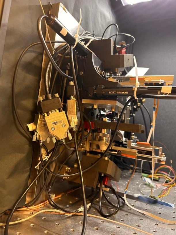
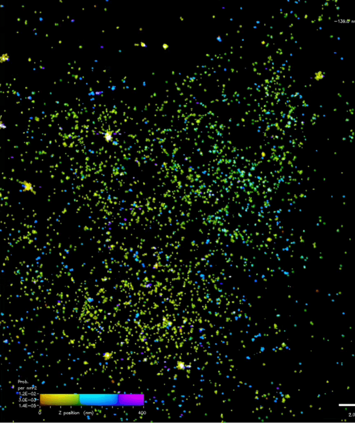
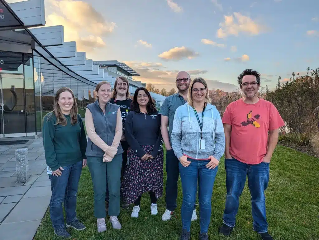
In November 2024 a group from the Kevin Hamill lab (https://lantsandlaminins.com/) at the University of Liverpool (UK) visited the AIC for 3 weeks to image components of the basal cell membrane using interferometric PALM (iPALM). The AIC iPALM system can image at a resolution of under 25 nm not only in x and y as with conventional PALM systems but can also reach up to 15 nm resolution in z. This makes it uniquely suited to image protein localization among filaments, the plasma membrane and other structures close to the cell surface, like focal adhesions and stress fibers. More information about the system can be found on the AIC website: https://www.aicjanelia.org/ipalm
The group around Kevin consisted of PhD student Natasha Chavda, Imaging Specialist Thomas Waring from the University of Liverpool Centre for Cell Imaging, and lab alumni Lee Troughton (now an Assistant Professor at Loyola University Chicago). This group applied to the AIC to investigate the 3D organization of Laminins in cells forming adhesions to the surface.
In the rest of this post, Kevin describes their visit and a typical imaging day.
As we sit at Washington Dulles airport waiting for the first leg of our trip home it seems fitting to reflect on what has been an intensely enjoyable, eye-opening, exciting, at times frustrating, but overwhelmingly awesome journey of discovery using the iPALM microscope at HHMI Janelia. Best ‘scope in the world, best three weeks of science-ing in my career.
Like with most experiments, once you get something to work you ‘crank the handle’ and try to maximise the output. This is especially true when you have finite access to the equipment. For the final week of our 3-week visit, Natasha Chavda and I were joined by LaNtsandLaminin alumni Lee Troughton, who flew in from his current position at Loyola, Chicago to help with handle cranking and to replace, Tom Waring who could only join us for two weeks.
We now travel back from our visit with data from 21 regions of interest. That sounds like a tiny amount (but 21 cells are >5 million individual images, ~7 TB of data) for people used to conventional microscopy, so I thought it would help giving an idea of the work required to acquire our data.
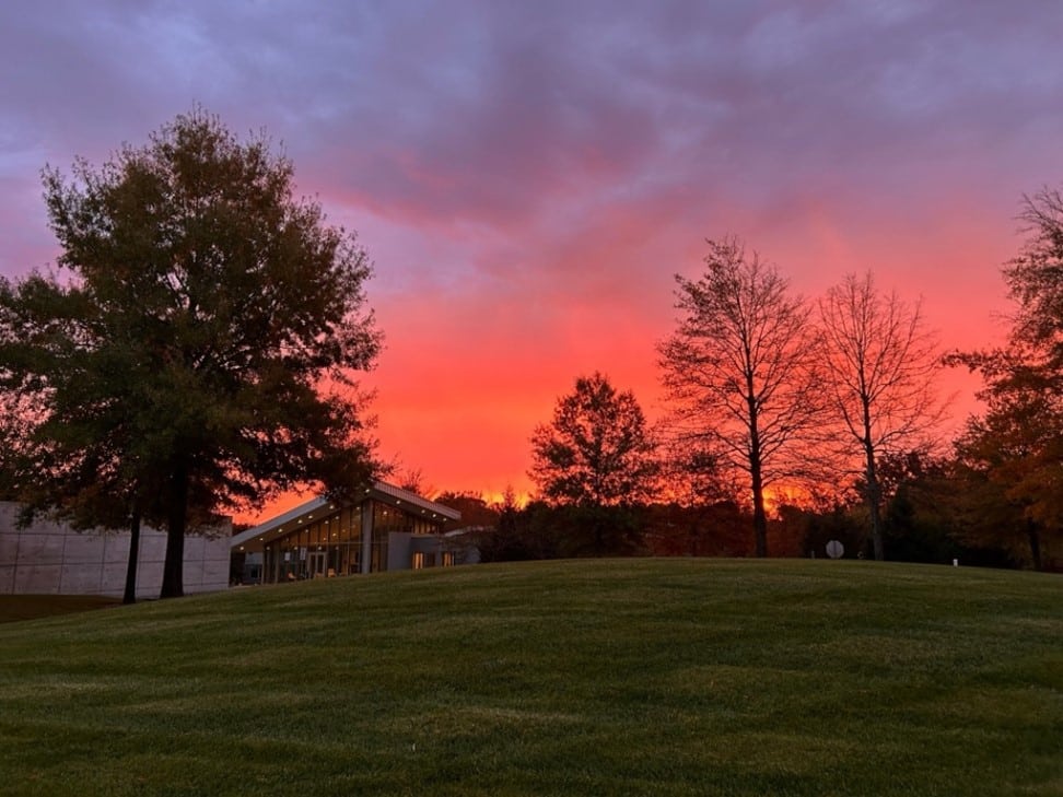
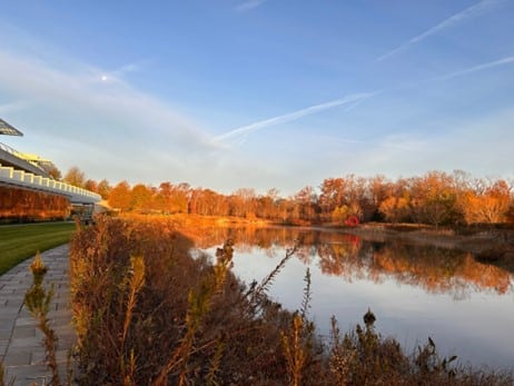
Our standard day started around 6am with me or Lee starting to process the samples. Antibodies were on before 7 am, then we would head down to Janelia’s restaurant ‘Bob’s’ for breakfast (or outside for a ‘golden hour’ pic of the pond). By 9 am the coverslips for the day were ready for Anja Schmidt from the AIC to add imaging buffer, seal and load onto to the ‘scope.
Around 9.15 am we selected the first cell to image., Then Anja began the initial alignment; first twiddling some tiny screw drivers to align the two objectives; correcting the final XY alignment and focusing with minute adjustments; adjusting camera/beam splitter location; then tweaking the beam splitter mirror so the correct quantity of light would reach each camera. We’d then wait for the system to settle and repeat the whole process again! After running a calibration run, we’d finally hit go on the first image by around 11 am. During this time, we would be finishing up the processing the images from the night before.
We’d have an early lunch while the 100,000 frames (~1 h) were collected in the 640 nm channel, then we’d have to repeat the process of alignment adjustments for the second channel. While the next 150,000 images (2.5 h) were collected in the 561 nm laser line, we ran the calibration, the initial macro and started the post-acquisition corrections for tilt and drift.
Around 3-3.30 pm we would be ready to start on the second cell. Choosing a good specimen and we’d start the alignment process, while we also started processing the 540 nm channel. By about 7 pm image acquisition for both channels for cell two would be complete.
Between 7-8 pm Natasha would be working in the cell culture hood preparing the cells for the next day’s imaging and also maintaining the multiple other lines needed for our project. Thereafter we would get going with the processing steps and macro running of the cell two data, so it was ready for analysis in the morning.
In the last few days, Lee and I were trained up on how to set the imaging up ourselves. By the end of the day the system was suitably settled that the alignment steps were achievable by us (mostly…). We did require Anja to come back in to help us at 8 pm on the first day and a technical consultation day 2 and 3. To help us with this process we created a reference video filming Anja doing all the steps, but it’s not easy to get right! So, by 7-9 pm (it took us substantially longer to align than the pro) we were able to image cell 3 channel 1 and, eventually, cell 3 channel 2. Our imaging acquisition finished after midnight. Extra data = extra processing, and by the middle of the final week Natasha would be spinning between three computers getting things going into the early hours.
We’re going home now pretty exhausted, but incredibly satisfied.
I am extremely happy that I was able to bring multiple people with me. It was definitely a team effort. Natasha was splitting cells every day while Tom, Lee and I stained coverslips. Everyone pitched in to analyse the data. Without the team, we’d either be zombies by now, would not have processed as much of the data and certainly wouldn’t have gathered as much data in the first place.
However, despite doing as much data processing as we possibly could during our trip, it is clear that there is so much more to do. Most of the things we’ve done so far are about quality control, but that only really gets us to the point where we can begin to answer the biology.
If you want to read more about our iPALM adventure or LaNts and Laminins, you can visit our lab blog: https://lantsandlaminins.com/
 (No Ratings Yet)
Get involved
Create an account or log in to post your story on FocalPlane.
More posts like this

Filter by
- NewsApply
- DiscussionsApply
- How toApply
- ToolsApply
- Case studiesApply
- InterviewsApply
- JobsApply
- EducationApply
- Blog seriesApply
- Latin America Bioima..gingApply
- From Zero to Qupath ..HeroApply
- Asian Microscopists ..and Cell BiologistsApply
- AIC at HHMI JaneliaApply
- Deep Learning for Bi..o-image analysisApply
- GloBIAS – updates fr..om the communityApply
- Volume EMApply
- Latin American Micro..scopistsApply
- Bio-image Analysis w..ith NapariApply
- Imaging with…Apply
- Towards Global Acces..sApply
- Highlights from Euro..-BioImagingApply
- LSFM seriesApply
- DIY MicroscopyApply
- View all