Microscopy preprints – applications in cell biology
Posted by FocalPlane, on 19 December 2022
Here is a curated selection of preprints published recently. In this post, we focus specifically on preprints using microscopy tools in cell biology.
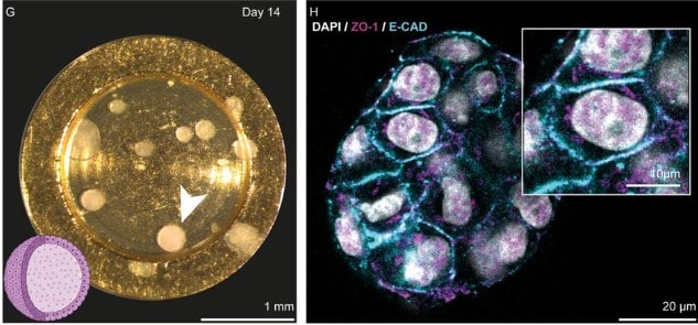
Light and electron microscopy continuum-resolution imaging of 3D cell cultures
Edoardo D’Imprima, Marta Garcia Montero, Sylwia Gawrzak, Paolo Ronchi, Ievgeniia Zagoriy, Yannick Schwab, Martin Jechlinger, Julia Mahamid
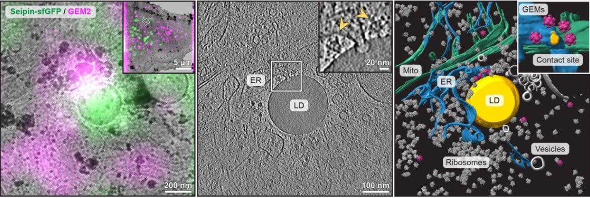
Genetically encoded multimeric tags for intracellular protein localisation in cryo-EM
Herman KH Fung, Yuki Hayashi, Veijo T Salo, Anastasiia Babenko, Ievgeniia Zagoriy, Andreas Brunner, Jan Ellenberg, Christoph W Müller, Sara Cuylen-Haering, Julia Mahamid
Tissue-Like 3D Standard and Protocols for Microscope Quality Management
Benjamin Abrams, Thomas Pengo, Tse-Luen Wee, Rebecca C. Deagle, Nelly Vuillemin, Linda M. Callahan, Megan A. Smith, Kristopher E. Kubow, Anne-Marie Girard, Joshua Z. Rappoport, Carol J. Bayles, Lisa A. Cameron, Richard Cole, Claire M. Brown
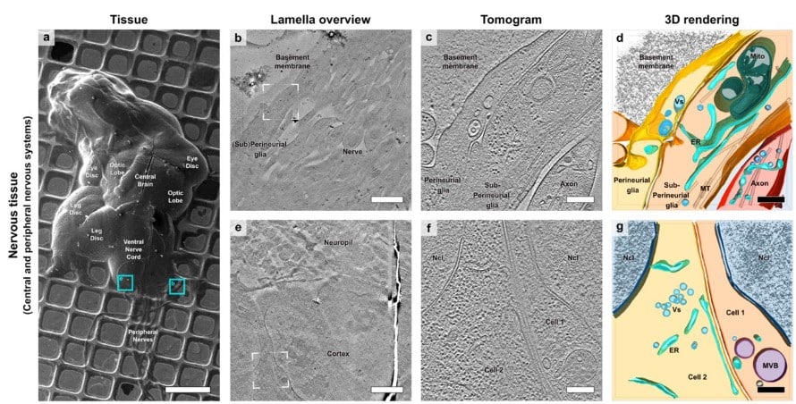
Cryo-electron tomography of native Drosophila tissues vitrified by plunge freezing
Felix J.B. Bäuerlein, José C. Pastor-Pareja, Rubén Fernández-Busnadiego
Multifocal two-photon excitation fluorescence microscopy reveals hop diffusion of H-Ras membrane anchors in epidermal cells of zebrafish embryos
Radoslaw J. Gora, Redmar C. Vlieg, Sven Jonkers, John van Noort, Marcel J.M. Schaaf
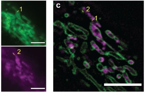
Long-term, super-resolution HIDE imaging of the inner mitochondrial membrane in live cells with a cell-permeant lipid probe
Shuai Zheng, Neville Dadina, Deepto Mozumdar, Lauren Lesiak, Kayli Martinez, Evan W. Miller, Alanna Schepartz
Organelle-specific photoactivation and dual-isotope labeling strategy reveals phosphatidylethanolamine metabolic flux
Clémence Simon, Antonino Asaro, Suihan Feng, Howard Riezman

Designed sensors reveal normal and oncogenic Ras signaling in endomembranes and condensates
Jason Z. Zhang, William H. Nguyen, John C. Rose, Shao-En Ong, Dustin J. Maly, David Baker
Direct Cryo-ET observation of platelet deformation induced by SARS-CoV-2 Spike protein
Christopher Cyrus Kuhn, Nirakar Basnet, Satish Bodakuntla, Pelayo Alvarez-Brecht, Scott Nichols, Antonio Martinez-Sanchez, Lorenzo Agostini, Young-Min Soh, Junichi Takagi, Christian Biertümpfel, Naoko Mizuno
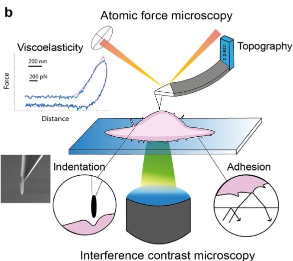
Coupled mechanical mapping and interference contrast microscopy reveal viscoelastic and adhesion hallmarks of monocytes differentiation into macrophages
Mar Eroles, Javier Lopez-Alonso, Alexandre Ortega, Thomas Boudier, Khaldoun Gharzeddine, Frank Lafont, Clemens M. Franz, Arnaud Millet, Claire Valoteau, Felix Rico
Time-resolved cryo-EM reveals early ribosome assembly in action
Bo Qin, Simon M. Lauer, Annika Balke, Carlos H. Vieira-Vieira, Jörg Bürger, Thorsten Mielke, Matthias Selbach, Patrick Scheerer, Christian M. T. Spahn, Rainer Nikolay
Single-molecule analysis of receptor-β-arrestin interactions in living cells
Jak Grimes, Zsombor Koszegi, Yann Lanoiselée, Tamara Miljus, Shannon L. O’Brien, Tomasz M Stepniewski, Brian Medel-Lacruz, Mithu Baidya, Maria Makarova, Dylan M. Owen, Arun K. Shukla, Jana Selent, Stephen J. Hill, Davide Calebiro
Live Imaging of Cutaneous Wound Healing in Zebrafish
Leah J. Greenspan, Keith Ameyaw, Daniel Castranova, Caleb A. Mertus, Brant M. Weinstein
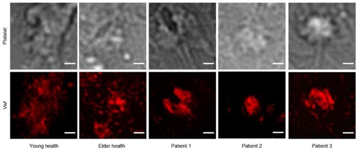
Changes of platelet morphology, ultrastructure and function in patients with acute ischemic stroke based on super-resolution microscopy
Bingxin Yang, Xifeng Wang, Xiaoyu Hu, Yao Xiao, Xueyu Xu, Xiaomei Yu, Min Wang, Honglian Luo, Jun Li, Yan Ma, Wei Shen
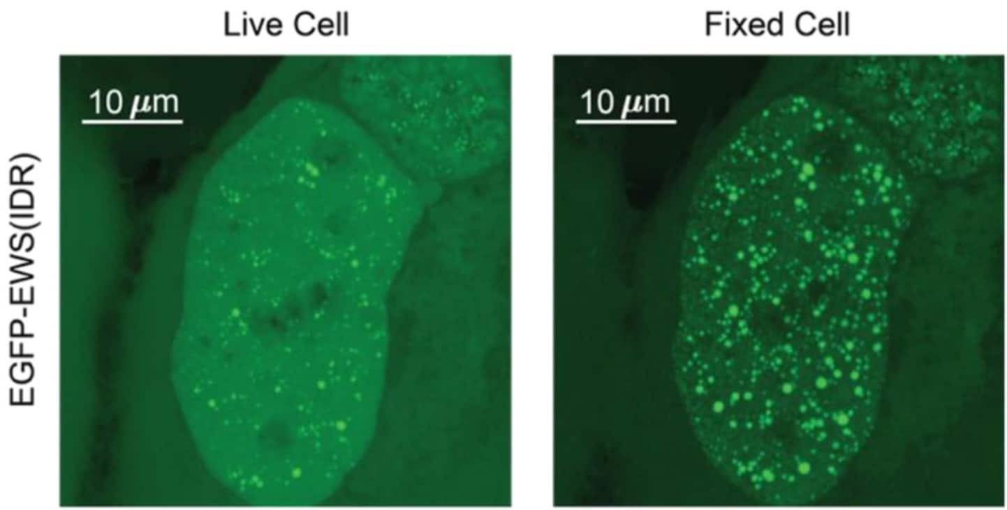
Fixation Can Change the Appearance of Phase Separation in Living Cells
Shawn Irgen-Gioro, Shawn Yoshida, Victoria Walling, Shasha Chong
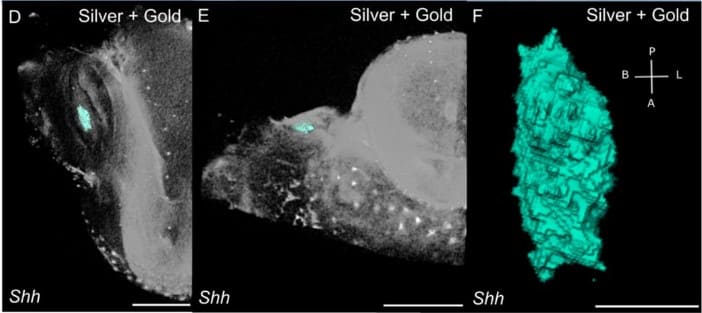
GECT – Gene Expression Detection in Developing Tissue Using μCT Imaging
Vilma Väänänen, Mona M. Christensen, Heikki Suhonen, Jukka Jernvall


 (No Ratings Yet)