Enhancing Global Access: interview with CZI grantee Beth Cimini
Posted by Mariana De Niz, on 24 January 2024

COLLABORATING ON CUSTOMIZED IMAGE ANALYSIS AND COMMUNITY ENGAGEMENT
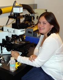
Beth Cimini is the Associate Director for Bioimage Analysis for the Imaging Platform at the Broad Institute of MIT and Harvard, where together with her team she directs efforts of software engineering including tools such as CellProfiler, researching morphological profiling and developing imaging assays. Her efforts also include a Postdoctoral Training Program in Bioimage Analysis, which aims to bridge an important gap in expertise in quantitative image analysis in academia and industry alike. She is a grantee of the second Cycle of CZI grants for imaging scientists.
What was the inspiration for your project? How did your idea for the CZI project arise?
Officially, what my CZI imaging scientist project was, was the Postdoctoral Training Program. The motivation happened during my own career. In joined Anne Carpenter’s lab in 2016 to do image analysis as a postdoc. She hired a lot of other postdocs too, to do image analysis, and many of them were formerly doing wet lab. For image analysis, expertise from both, wet lab and dry lab is valuable, because in our position, if the images aren’t of enough quality, we should be able to help the user figure out at what level the quality could improve. We cannot just say ‘this doesn’t work, do better’. Instead, we should be in a position to provide advice on both the sample preparation, the biology itself, image acquisition, and ultimately image analysis. So, we realized it was helpful to have biologists turned computer scientists, and we saw there were many people interested in this transition. We were inspired by Jennifer Waters’ fellows program. We had been providing training so far, and we realized we were doing a good job at it, so we wanted to formalize this. We spoke about doing this since 2017, and we hired the first group in 2019- and the CZI funded me a year later. So far, we have had 7 postdocs who have started and finished, and 4 who are currently in our team.
CellProfiler has been key for many microscopists worldwide. How does that fit into the equation of your project?
We are building image analysis workflows for other researchers as part of what we do. Almost nothing in CellProfiler is algorithmically novel, nor does it have special Maths that you can’t find in other types of software. What CellProfiler does, however, is taking these computer science advances and making them interpretable and usable by biologists. Having a team of postdocs that are fresh from the wet lab joining the dry lab is very valuable. They make tools that are useful for others and can give us ideas of what we can do better, based on experience they might have acquired in their PhDs or other postdocs. They have made incredibly important contributions to CellProfiler, some on the code level- those who want to develop software engineering skills; some who give ideas in terms of usability and how we can improve our work; and some who can teach people how to apply this to their work.

To you, what is the importance of community engagement for advancing microscopy? And what is the importance of language in bridging gaps and facilitating communication between different areas of expetise?
I think facilitating communcation between scientists across disciplines is everything. If we stay ‘siloed’ in our individual labs or our individual sub-disciplines, science doesn’t grow. In our lab we are lucky to have people from multiple backgrounds including physics, computer science, mathematics, biology, etc. There are so many similar problems being solved in individual siloes out there, that the waste of time and resources is regrettable. It makes me sad how much time is wasted re-inventing the wheel in 100 different labs. We try to address this with CellProfiler: people might be trying to figure ways to analyse histones or telomeres, but at the end of the day they are speckles inside a nucleus. We should be using the same solutions. Our aim is to help people realize that they might share similar problems, and facilitate communication between the to share solutions to problems. We could be devoting our energy to making a million new things, instead of millions of copies of the same thing.
There aren’t a lot of people doing what you are doing. Where do you see your project in 10 years?
I hope that I will still be solving image analysis problems in 10 years, just different ones. For instance, from a computer science perspective, segmentation is almost a solved problem. We are very close to having a generalizable solution that if you have a computer science degree and access, you can devleop segmentation for just about anything within a couple of hours. That’s not yet true for biologists. So I hope in, say, 5 years we are spending less time helping people do segmentation, unlike now, when we spend 99% of our time on this. I hope I will still be solving interesting problems and helping people close the gap. I don’t think the need for image analysis is going to decrease. Even if segmentation were magically solved tomorrow, there would still be a need for image analysis in terms of measurements and more complex questions. Maybe in 25 years all our current problems will be solved, but my guess is microscope developers will come up with new cool things for those of us who are doing image analysis to analyse!
How did you first hear about CZI, and how has their support helped you reach your aims?
The CZI funding totally enabled what we are doing. It allowed for the flexibility that is neede when people switch fields: people don’t need to know exactly what they are doing right from the start. They can take a couple of months to get up to speed. Allowing people to learn at the correct speed is so valuable. I don’t remember where I first heard about CZI, other than when it was originally announced. A lot of the open source image analysts and software devleopers came together and organized a meeting where we wrote this open letter to them stating the importance and need of open software and image analysis for the advancement of science. I don’t know if that letter had any impact- I imagine it did because even before they started the imaging scientists program, they started the imaging software fellow program where they funded developers from different open tools, including CellProfiler. This funding was really unique because while it’s “easy” to obtain NIH funding to build new software, getting funding to support and maintain software that already exists but is not novel is basically impossible.

How can we as an imaging community foster cross-disciplinary communication and engagement to connect disciplines the way you have so successfully done so?
I think there is still a lot of fear towards or hesitance, or even annoyance about having to do image analysis. Learning to do something new is scary and hard. But in the same way that we understand that when we join a lab that does microscopy, we are going to have to learn to do sample preparation, and how to use different microscopes, we should realize that image analysis is not an option or something detached. It should be as organic as learning how to properly operate a microscope. The imaging community has to get on board with the fact that image analysis is part of the cycle.
In what ways has your project democratised microscopy and increased access?
Everything we do is open source and free, and we try to be as accessible as possible. This includes our new web-based tool, piximi, which can run from a browser in your phone or in an old kindle. We hope we can democratize analysis in that way. From our postdoctoral program, two amazing members, Barbara Diaz-Rohrer and Rebecca Senft led a paper that we published in PLOS Biology, titled ‘A biologist’s guide to planning and performing quantitative bioimaging experiments’. They coordinated with several authors from the BINA Training and Education and Image Informatics Working Groups, and made this resource available. We hope this helps users in providing a starting point: if you are at a place where you have access to a great core, and a great core director and staff who can teach you all the things you need to learn, that’s great. If you don’t, how do you even know what you need to learn? How do you know what you don’t know, and what the critical skills are? We hope this paper is a (poor) substitute, but still a substitute for the missing core. In addition, we have created a companion Jupyter book at bioimagingguide.org where we are collecting resources. While the paper is a snapshot in time, this can be a living resource, explicitly aimed at beginners. We have also started working with BINA and other groups to start translating this into other languages. We have teams working on 7 different languages now. We want to assemble and develop good free educational resources, we want to make sure that people can find them, and that they are in a language that they speak and understand.
What challenges have you faced during the creation of your project, and what have been your biggest successes?
Our biggest success are the 7 amazing humans who have gone through the postdoctoral program and have now moved on to jobs they are happy and excited about. I could not be more proud. Regarding challenges, getting people to pay for image analysis is hard, and getting people to fund open source is hard. We run a research group and a core facility at the same time, so there are lots of challenges in balancing those things: how much time we spend developing new software versus helping people with existing ones. At the end of the day, we are grateful for the support for this project, and for the work we have been able to do. We hope now we have made a model that other places will see as valuable and want to follow. We think training programs (postdoctoral, internship and apprenticeship programs), when done thoughtfully and carefully, avoiding exploitation and really thinking about what the trainee is going to learn, can be extremely powerful. For not so much money, they can result in a tremendous amount of expertise and scientific cross-collaboration. We hope this work continues to be catalytic, and that we can keep doing it.
Check out our introductory post, with links to the other interviews here


 (No Ratings Yet)