Featured image with Rosa L. Salgado García
Posted by FocalPlane, on 6 June 2025
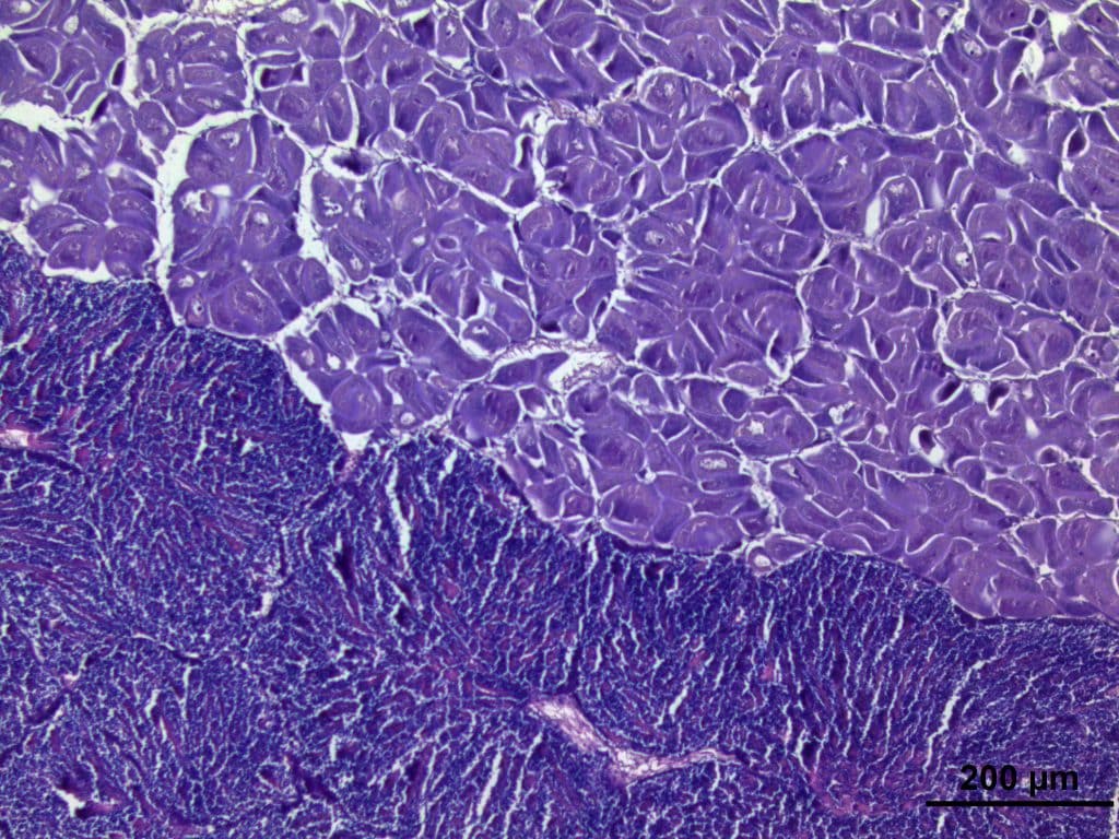
Our featured image, acquired by Rosa L. Salgado García, shows a histological section of female (above) and male (below) gonads at an advanced maturation stage of the simultaneous hermaphroditic scallop Nodipecten subnodosus. The image of the gonadic tissue was obtained using a microscope (Leica DM4B) connected to a digital camera (Leica, DMC2900) using LAS V software 4.12 at the Centro Interdisciplinario de Ciencias Marinas (CICIMAR IPN). Each gonad portion was dehydrated in alcohol at increasing concentrations (70–100%), cleared with Hemo-De and embedded in Paraplast-Xtra. 4 μm-thick sections were cut on a microtome and stained with Harris hematoxylin and eosin.
Discover more about Rosa’s research
Research career so far: I currently work in the Energy Metabolism lab in the Aquaculture Department from the Centro de Investigaciones Biologicas del Noroeste (CIBNOR). During my PhD thesis, I investigated the metabolic responses of scallop N. subnodosus broodstock exposed to hyperthermia during reproductive effort. My objectives focused on understanding the response of animals facing hyperthermia, as high mortality was observed during summer, when animals simultaneously face high energy demand for reproduction and temperatures beyond the thermal tolerance window of species. For this, I identified the reproductive condition of scallops by analyzing the histology of gonads using microscopy. Then, I analyzed the energy content (ATP, adenosine triphosphate) by HPLC and the activity of enzymes that participate actively in cell energy metabolism. Currently, our research group collaborates with researchers from other institutions like the University of Brest (UBO, France) to decipher the thermal biology of N. subnodosus integrating proteomic, genetic, and metabolism data obtained from different populations from the North Pacific Coast of Baja California Sur, Mexico.
Current research: My research focuses on understanding the energy metabolism involved in resilience and the adaptative capacity of marine animals in the context of climate change. By using microscopy, we provide confident data about the quantity and quality of cells, such as gametes, providing pivotal information on the animal’s capability to allocate energy to survive and adapt to changing environments, allowing us to make better decisions for the conservation and management of marine ecosystems.
You can find more information about Rosy Salgado research work here: https://www.cibnor.gob.mx/investigacion/acuicultura/pa-laboratorios/metabolismo-energetico
Favourite imaging technique/microscopy: I enjoy using light microscopy and quantitative imaging techniques, and I would also like to learn about fluorescence microscopy imaging techniques to study mitochondria metabolism.
What are you most excited about in microscopy? I am excited about the potential of microscopy to identify animals’ physiological responses to environmental drivers within marine ecosystems. Microscopy offers insights into animal physiology, enabling us to explore the complex mechanisms in cells that underpin survival and adaptation of animals coping with challenging environmental conditions.
 (1 votes, average: 1.00 out of 1)