Imaging with A*STAR Microscopy Platform
Posted by FocalPlane, on 15 October 2025
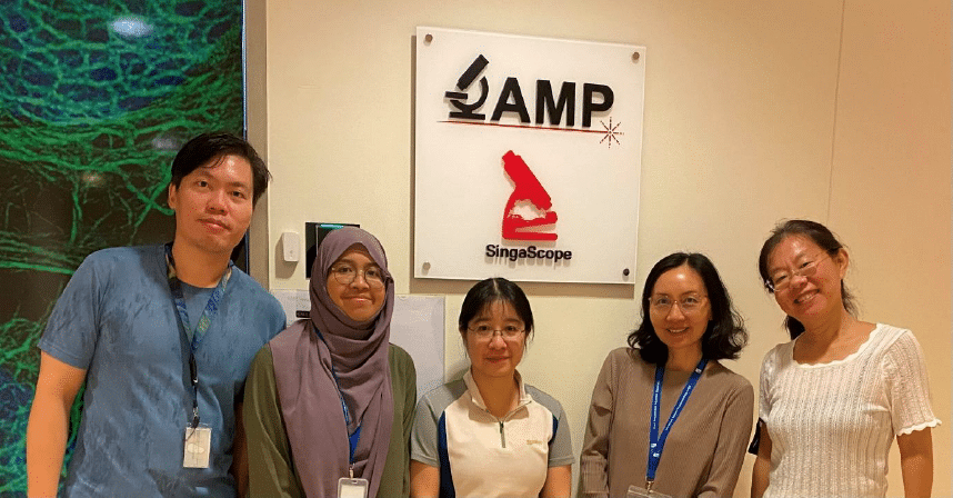
In our ‘Imaging with…’ blog post, we meet the staff of the A*STAR Microscopy Platform in Singapore.
Staff role call
Dr. MA Xiaoxiao, Manager
Expertise: Knows a bit about everything in the facility, and familiar with a few things in microscopy!
Most likely to be found struggling to allocate time among training sessions, reports, meetings (including preparing them), and planning.
Dr. GOH Wah Ing, Assistant Manager
Expertise: Light microscopy, experimental design, firefighting required for facility and lab management.
Most likely to be found cleaning objective lenses and disassembling microscopes to salvage spare parts.
John LIM, Image analysis expert
Expertise: Image analysis, light-sheet microscopy & part-time IT
Most likely to be found either at my laptop or at the image analysis computer
Annisa IBRAHIM, Research Officer
Expertise: Histological slide scanning, live cell imaging and widefield fluorescence
Most likely to be found in front of the slide scanner setting up and optimising scan profiles
Dr. LIN Shuping, industry collaborator who literally stays with us forever in the facility
Expertise: Confocal microscopy and live cell imaging
Most likely to be found helping users or training users at microscopes.
Microscope role call:
Leica Stellaris 8 Falcon FLIM, LaVision Trim II multiphoton (with both upright and inverted microscopes), Yokogawa X1 spinning disk confocal plus 3D FRAP plus Live-SR SIM, Zeiss LSM880 Airyscan, Miltenyi UM II lightsheet, ONi STORM, Zeiss Axioscan 7 (THE money maker), a bunch of great workhorses such as Evident FV3000RS and Zeiss LSM800 scanning confocals, and very useful brightfield and widefield systems (upright and inverted live cell imaging microscopes)
Who can access the facility?
Anyone who’s willing to undergo the training and be responsible enough to keep the facility clean and organised for everyone to enjoy.
Pet peeve (something that users do that is annoying): Consistently leaving the microscope in a mess and disregarding our shutdown instructions—plus getting oil on air lenses and focus knobs!
Favourite microscope: Everyone has her/his own favourite microscope! We don’t want to start a fight… but, we have come to an agreement that the favourite is the best one for answering the targeted scientific question.
Favourite thing to image: Whenever something new that we have never imaged before comes in, there is excitement everywhere!
Best bit of advice (that you give or have been given)
In front of the microscopy: One drop of oil is all you need—no more, no less!
For all experiments, (especially for PhD students in despair): Today is the first day of the rest of your life.
If money was no object, we would buy… Our imaginations ran wild—think high-speed data transfer networks and a dream team of extra facility staff (though “buying” them might not be the best phrasing… but you get the point!). Back in reality, our more achievable wish list includes upgrading all FV3000s to FV4000s and the LSM800 to LSM900/980—because these machines are absolute workhorses that refuse to quit. Oh, and if we could also get something that does everything our beloved 2-in-1 multiphoton microscope and spinning disk confocal, we would be more than happy!
Can you give us some examples of recent papers that were published with your assistance?
Para-Hydroxycinnamic Acid Mitigates Senescence and Inflammaging in Human Skin Models. DOI:10.3390/ijms25158153
This is a great example of how we support researchers, with multiple staff contributing from different angles. Wah Ing guided them in selecting the right microscope(s) for different aspects of their scientific questions. Both Shuping and Wah Ing provided training, enabling the researchers to work independently on their project. When it came to post-image acquisition, John stepped in, offering his expertise in image analysis—earning him a spot in the authorship. A fantastic team effort!
How should users acknowledge the facility and why is it important?
The best way to recognise our contributions is by mentioning us in the publications, either as co-authors or in acknowledgements since this helps highlight our role in supporting research. We are not and should not be a profit-driven service but rather a support hub for sharing knowledge and expertise in light microscopy, benefiting the entire research community. With our experience, researchers save time, reduce costs in microscopy experiments, and avoid unintentional mistakes that could lead to incorrect conclusions or even image integrity issues. However, the value of knowledge sharing, isn’t always immediately visible, making recognition in publications essential to showcase our impact.
That said, we also deeply appreciate more tangible forms of feedback—like chocolates and desserts! Beyond just being delicious, they reassure us that users truly see us as friendly and helpful, which is just as rewarding.
 (1 votes, average: 1.00 out of 1)
Get involved
Create an account or log in to post your story on FocalPlane.
More posts like this

Filter by
- NewsApply
- DiscussionsApply
- How toApply
- ToolsApply
- Case studiesApply
- InterviewsApply
- JobsApply
- EducationApply
- Blog seriesApply
- Towards Global Acces..sApply
- Latin America Bioima..gingApply
- From Zero to Qupath ..HeroApply
- Asian Microscopists ..and Cell BiologistsApply
- AIC at HHMI JaneliaApply
- Deep Learning for Bi..o-image analysisApply
- GloBIAS – updates fr..om the communityApply
- WAMBIAN: West Africa.. in FocusApply
- Volume EMApply
- Latin American Micro..scopistsApply
- Bio-image Analysis w..ith NapariApply
- Imaging with…Apply
- Highlights from Euro..-BioImagingApply
- LSFM seriesApply
- DIY MicroscopyApply
- View all