Workshop report: York Confocal Microscopy course
Posted by Yelena Ivanova, on 3 February 2026
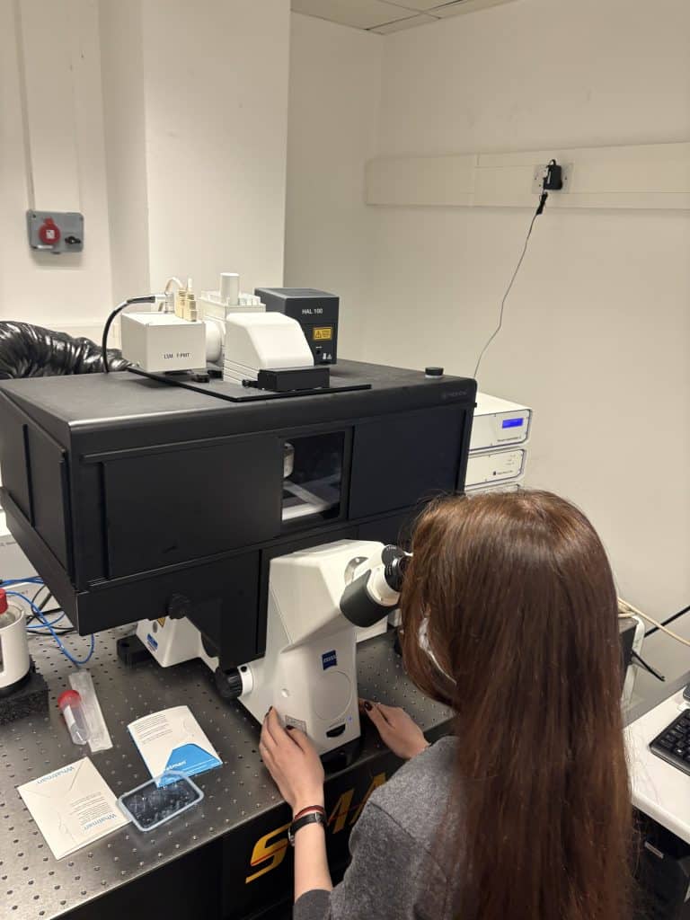
During the four-day Confocal Microscopy 2025 course at the University of York, I gained a better understanding of both the fundamentals of microscopy and more advanced techniques. The daily schedule consisted of the introduction of the relevant topics in the morning, followed by several hands-on practical sessions. In these, we were split into small groups and rotated across different confocal systems, allowing us to work with a variety of microscopes and imaging setups. As practice material, we were provided with a wide range of samples (from plant tissue to live cells) with different fluorescent labels. Furthermore, we had the opportunity to work with multiple demonstrators with different research interests and areas of expertise.
Previously, my experience with confocal microscopy was limited to three-colour imaging of fixed tissue. During the course, I learned and successfully used FRAP, FRET, and other live cell imaging techniques. These approaches are highly relevant to my research, as analysis of protein-protein interactions and trafficking dynamics would provide further detail into the mechanisms of Alzheimer’s disease.
One particularly exciting aspect of the course was the hands-on experience with spectral imaging with linear unmixing, which allows for the analysis of multiple fluorophores with overlapping spectra. Being able to visualise different tau species within the neuron would provide important information on the differences in tau localisation, misfolding, and aggregation. Another highlight of the course was the exploration of different strategies for improving the quality of the images. Capturing fine neuronal processes is crucial for neurodegeneration analysis, and learning how to use new software-based techniques was very rewarding.
During the practicals led by industry representatives, we operated both laser scanning and spinning disk confocal systems and discussed their respective advantages and limitations in terms of resolution, speed, and flexibility. Furthermore, we observed and compared different ways to significantly decrease imaging time, which would allow for more rapid data collection without sacrificing image quality. Importantly, we also had an opportunity to test different image optimisations on our own samples.
Overall, this course improved my understanding of the theory behind confocal microscopy and allowed me to use cutting-edge techniques that will directly benefit my research. Gaining hands-on experience with live cell functional imaging (FRAP, FRET) has significantly increased my confidence in applying these methods to study protein dynamics in Alzheimer’s disease. The practical aspect of the course, combined with the guidance from academia and industry experts, has equipped me with the skills to better design and conduct imaging experiments. I am grateful for the support that allowed me to attend this course and look forward to integrating everything I have learned into my current and future work.
 (3 votes, average: 1.00 out of 1)
Get involved
Create an account or log in to post your story on FocalPlane.
More posts like this

Filter by
- NewsApply
- DiscussionsApply
- How toApply
- ToolsApply
- Case studiesApply
- InterviewsApply
- JobsApply
- EducationApply
- Blog seriesApply
- Deep Learning for Bi..o-image analysisApply
- GloBIAS – updates fr..om the communityApply
- WAMBIAN: West Africa.. in FocusApply
- Volume EMApply
- Latin American Micro..scopistsApply
- Bio-image Analysis w..ith NapariApply
- Imaging with…Apply
- Towards Global Acces..sApply
- Latin America Bioima..gingApply
- From Zero to Qupath ..HeroApply
- Asian Microscopists ..and Cell BiologistsApply
- AIC at HHMI JaneliaApply
- Highlights from Euro..-BioImagingApply
- LSFM seriesApply
- DIY MicroscopyApply
- View all