FocalPlane image competition – vote for your favourite
Posted by FocalPlane, on 4 July 2023
Thanks to everyone who entered our 2023 image competition, we were delighted to receive so many wonderful entries. We have shortlisted 10 images for our public vote. You can vote using the poll at the bottom of this page and the image with the most votes will be featured on the cover of Journal of Cell Science and the scientist will win £100. Voting will close on Friday 4 August.
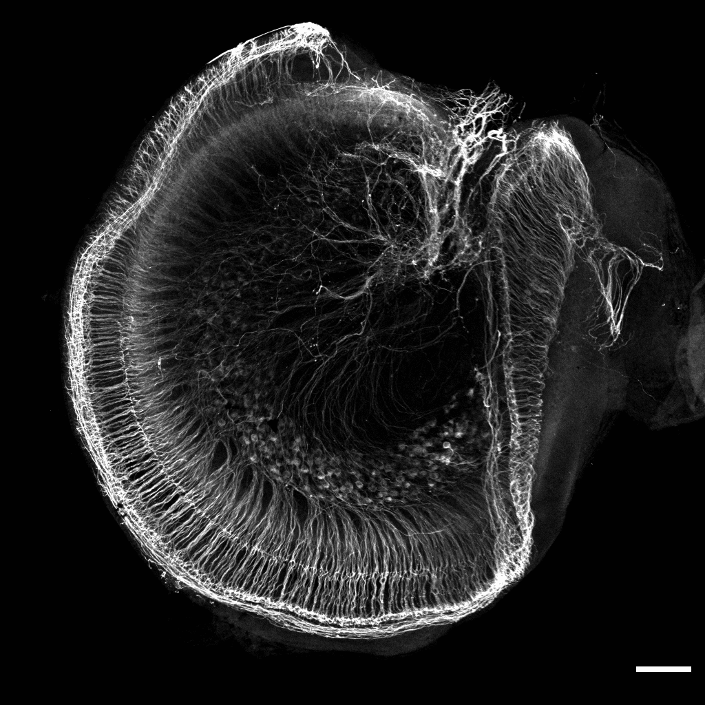
1. Spiral ganglion neuron explant – Raman Kaushik
P1 mice spiral ganglion neuron (SGN) explant cultured for 2 days and then stained with Tubb3 (gray) imaged at 10x air objective with LSM 780 Zeiss microscope. Image processed using ImageJ software.
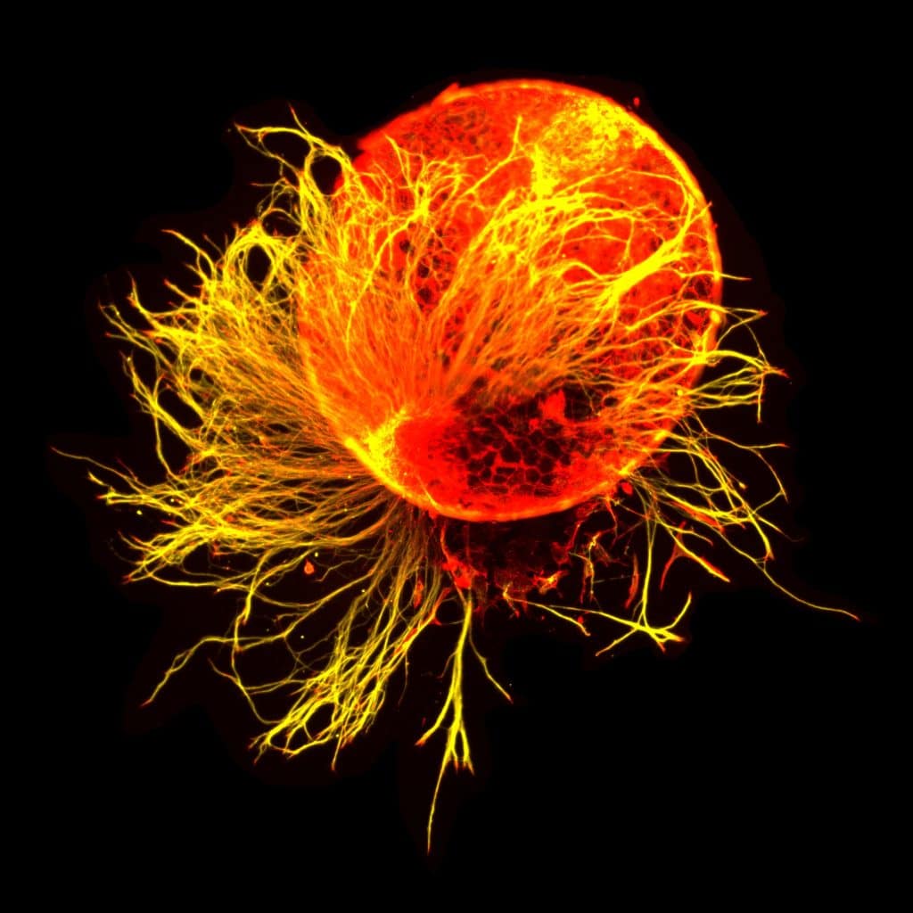
2. Xenopus retinal explants – Jana Sipkova
Xenopus retinal explants grown on glass for 24 hours, stained for tubulin (yellow) and actin (red). Imaged on a Leica DMi8 inverted microscope with 63×/1.4 oil immersion objective (ORCA-Flash4.0 V2 Digital CMOS camera Model C11440- 22CU). Post-processing was conducted in Fiji.
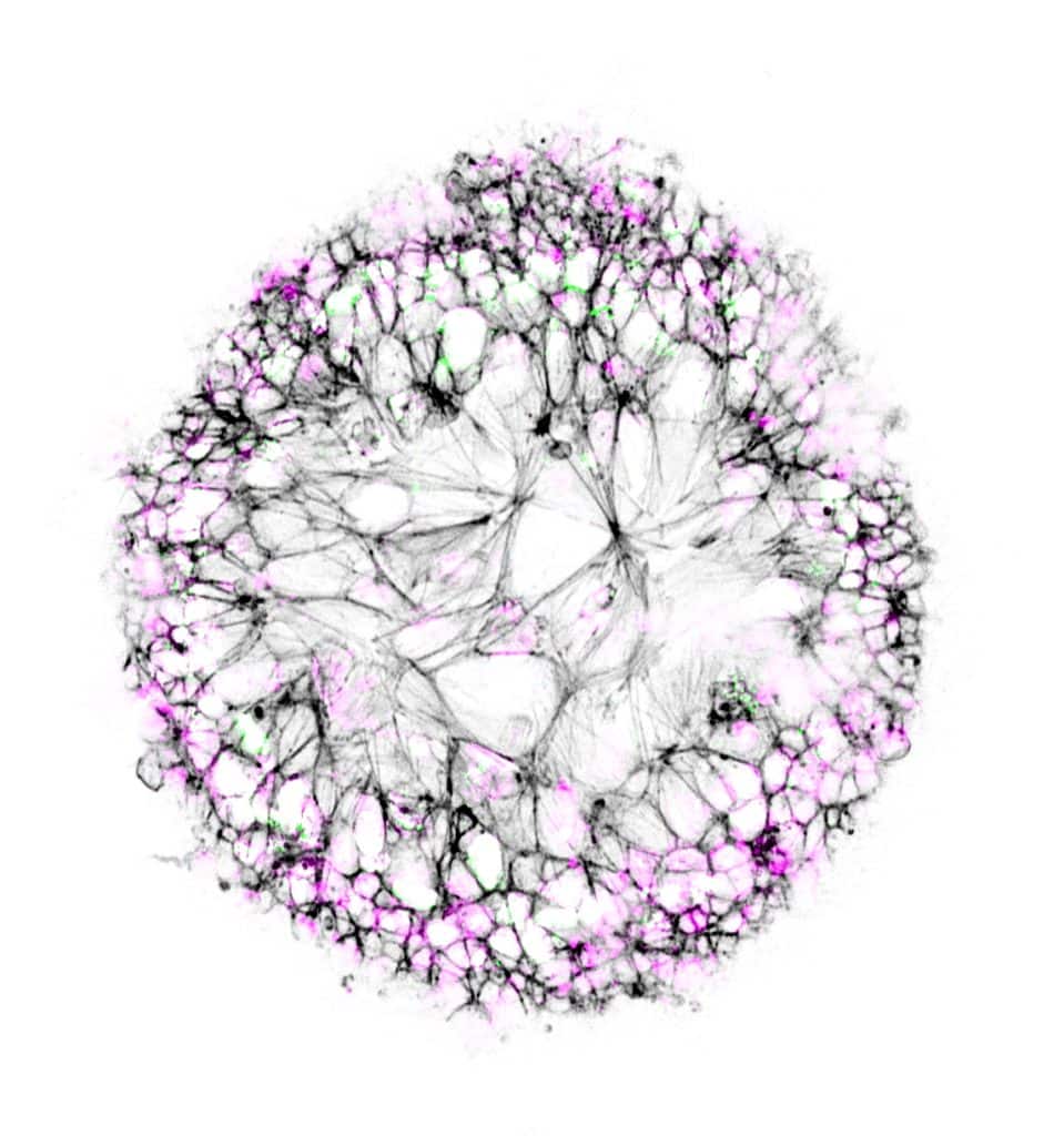
3. Dandelion in spring – Ioakeim (Makis) Ampartzidis
Neuroepithelial-like cells were cultured on glass beads. The cell nuclei are coloured in magenta, and the cell’s skeleton is stained against F-actin, in black. The actively proliferating cells are highlighted with Ki-67 antigen in green.
4. Primary mammary gland organoid – Oona Paavolainen
Primary mammary gland organoid in a collagen-I matrix formed from a single primary epithelial cell isolated from material obtained from a breast reduction surgery. Image was taken with a spinning disk confocal microscope (3i CSU-W1 Spinning disk) using a 20x Zeiss Plan-Apochromat (NA=0.8) objective. Constructed image was made in ImageJ and is a maximum intensity projection with a rainbow depth-coded lookup-table.
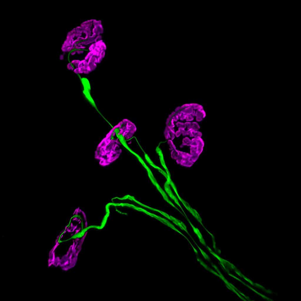
5. Neuromuscular junctions – Rebecca Simkin
Collapsed z-stack confocal image of neuromuscular junctions in a lumbrical muscle located in the hindpaw of a mouse. Lower motor neurons are visualised in green (2H3/SV2), and post-synaptic acetylcholine receptors in magenta (αBTX).
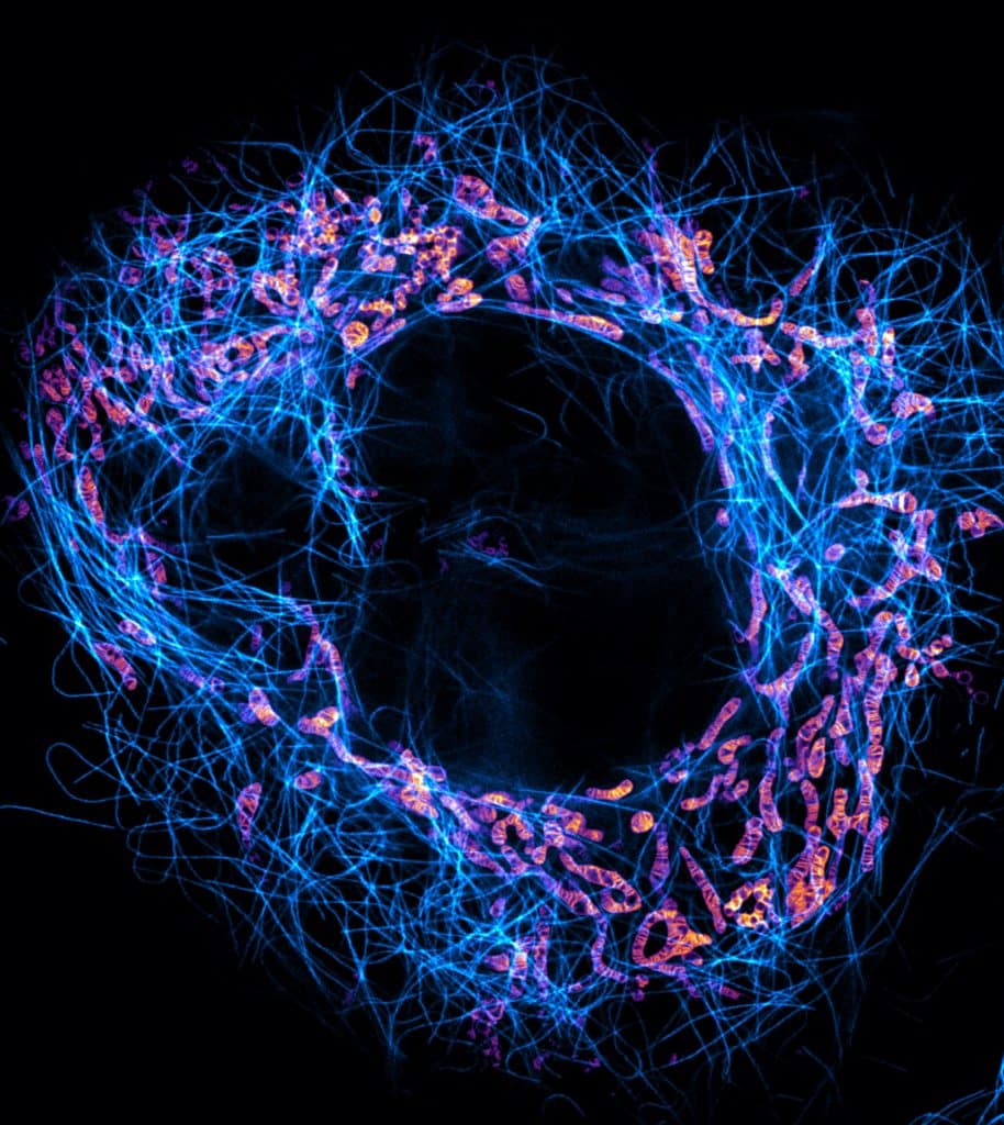
6. Microtubules and mitochondria – Till Stephan
The image shows a live-cell stimulated emission depletion (STED) microscopy recording of a HeLa cell in which mitochondria (orange-magenta) and microtubules (cyan) are labelled. The super-resolution image reveals the densely stacked mitochondrial cristae, coordinated invaginations of the mitochondrial inner membrane.
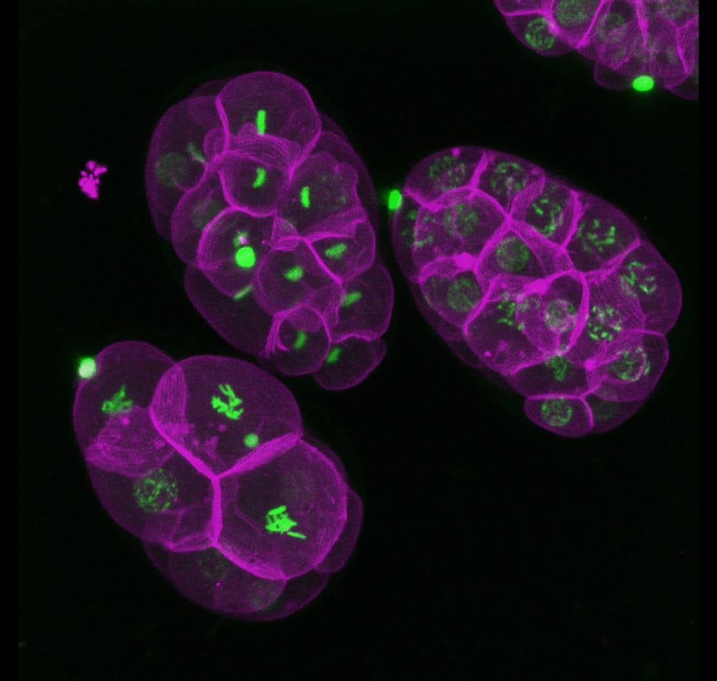
7. Transgenic C. elegans embryos – Martin Estermann
3D reconstruction of live imaging of C. elegans transgenic embryos. Embryos were imaged using a Zeiss Celldiscoverer 7 microscope. 3D reconstruction was performed using Fiji. Histones in green and cell membrane in magenta.
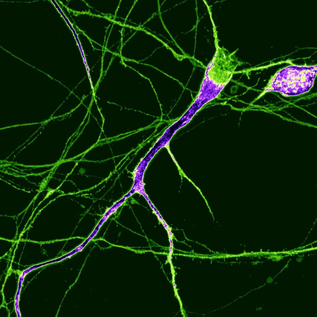
8. Dopaminergic neuron – Nick Gatford
This image shows a single human dopaminergic neuron generated from a human stem cell acquired via super-resolution Airyscan confocal microscopy at the University of Oxford Micron facility.
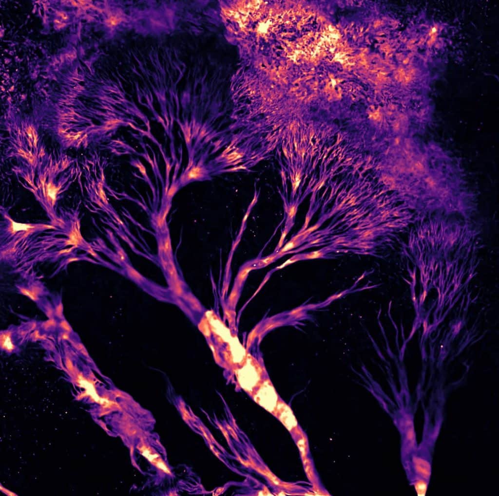
9. Dictyostelium Trees on Fire – Peggy Paschke
Dictyostelium discoideum cells stably expressing the cAMP sensor Flamindo2 were plated at high density and starved for 5 hours. Cells were imaged as a 5×5 tile scan with a Nikon AX Confocal microscope (488 nm excitation) using a 4x air objective for 5 hours. Shown is a cropped part of the last acquired tile scan, at the very edge of the agar slop, which creates the elution of Dicty Trees. Tile scans were stitched together in ImageJ. The LUT is mpl magma.
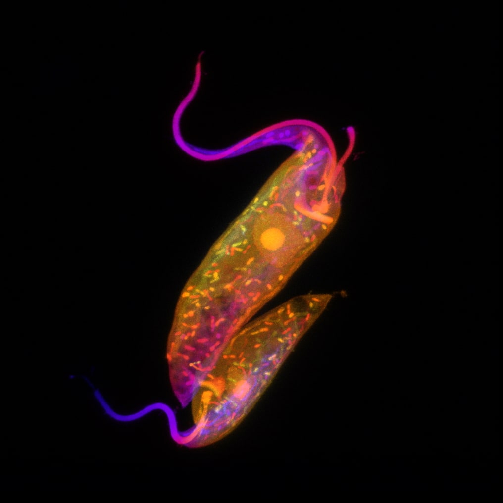
10. Trypanosoma cruzi epimastigote – Victoria Alonso
Trypanosoma cruzi epimastigotes prepared for expansion confocal microscopy, pan-proteome staining with NHS-ester Atto 594. Image acquired with Zeiss LSM 880, Z-stack. Image was edited with ImageJ Fiji, plugin: Z-stack depth Colorcode
Voting has now closed in our image competition.
Thank you for voting
 (37 votes, average: 1.00 out of 1)
43 thoughts on “FocalPlane image competition – vote for your favourite”
Leave a Reply
Get involved
Create an account or log in to post your story on FocalPlane.
More posts like this

Filter by
- NewsApply
- DiscussionsApply
- How toApply
- ToolsApply
- Case studiesApply
- InterviewsApply
- JobsApply
- EducationApply
- Blog seriesApply
- Volume EMApply
- Latin American Micro..scopistsApply
- Bio-image Analysis w..ith NapariApply
- Imaging with…Apply
- Towards Global Acces..sApply
- Latin America Bioima..gingApply
- From Zero to Qupath ..HeroApply
- Asian Microscopists ..and Cell BiologistsApply
- AIC at HHMI JaneliaApply
- Deep Learning for Bi..o-image analysisApply
- GloBIAS – updates fr..om the communityApply
- Highlights from Euro..-BioImagingApply
- LSFM seriesApply
- DIY MicroscopyApply
- View all
I love this image! The colours are beautiful.
All are very amazing images x
Well done to all of you x
Wow, I love number 5!
Great photo
Spectacular !
Love number 5
There is no structure more beautiful in biology than chromosomes.
Beautiful
The most impressive neurone staining I have ever saw
Fascinatingly beautiful
All are amazing but I pick this one due to its simplicity.
Good luck Becca!
All fabulous, however I am drawn to number 5
Love the simplicity
The number 7 is amazing
These are amazing… well done.
Good luck!
Fabulous Becca, well done xx
Wow what a stunning image who knew cells could look so beautiful good luck x
Amazing number 5.
So clever 👏🏻
Genial #7
El número 7 !!!
Number 5 looks like flowers!! Love x
Really beautiful!
Fantastic shot so vibrant
No5 … what a stunning array of cells … beautiful
Beautiful, well done
Number 5 – looks like sweet peas. Nice tattoo ❤️
Well done becca! It’s amazing what you do x
Love these images x
Beautiful images but the Dicty is gorgeous!!!
What a fitting name for #9 Trees on fire! such an amazing glimpse into a tiny space!
All amazing images, but I love the detail in number 9.
Number 9 is beautiful!
Beautiful images! I love the brilliant trees in Number 9!
Beautiful Dicty image (number 9)
Amazing images. Hard to pick but number 9!
The images are all beautiful! But I vote for #9 Dicty on Fire! Great work Peggy.
Spectacular work amazing.
Number 9 is phenomenally successful. Exzellent colors. Great work.👍
Todas las fotografías muy bellas. Gracias a los científicos que logran ver más allá del ojo humano y además “entienden” lo que están viendo. Voy por la 10!
Amazing x
Number 7
Martin Estermann