Advertisement

Displaying posts with the tag: is_archive

Microscopy preprints - applications in biology

Posted by , on 12 August 2025

Here is a curated selection of preprints published recently. In this post, we share preprints that use microscopy tools to answer questions in biology.

Featured image with Maik Bischoff

Posted by , on 18 July 2025

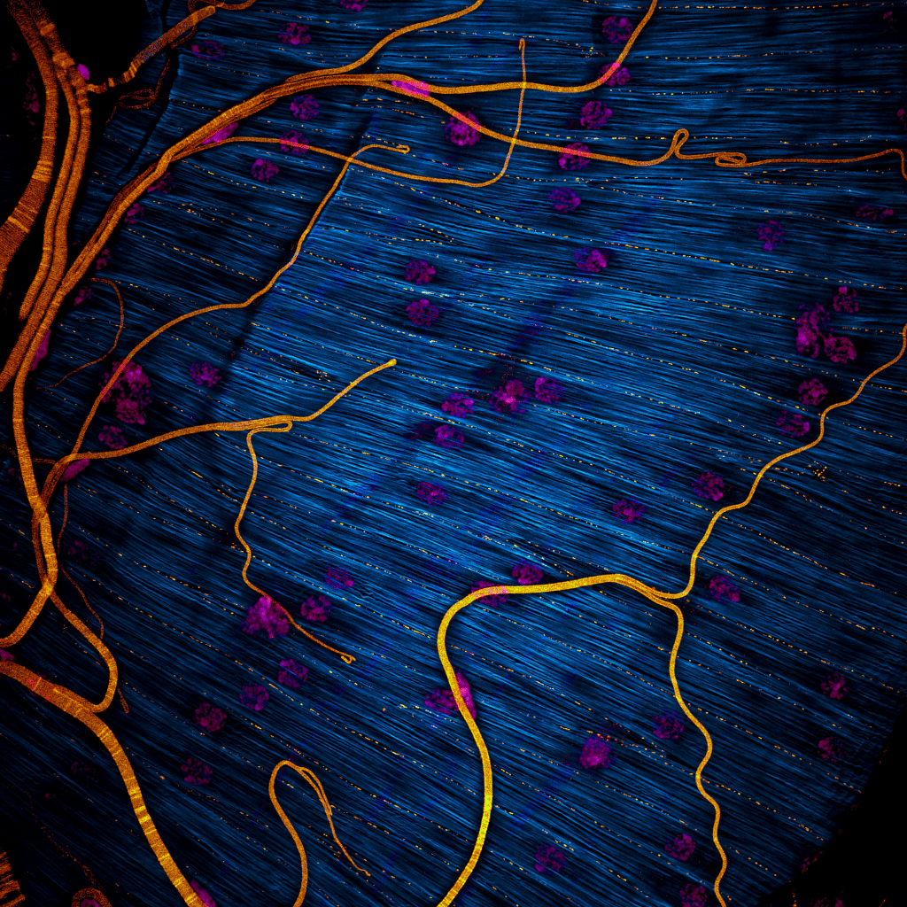
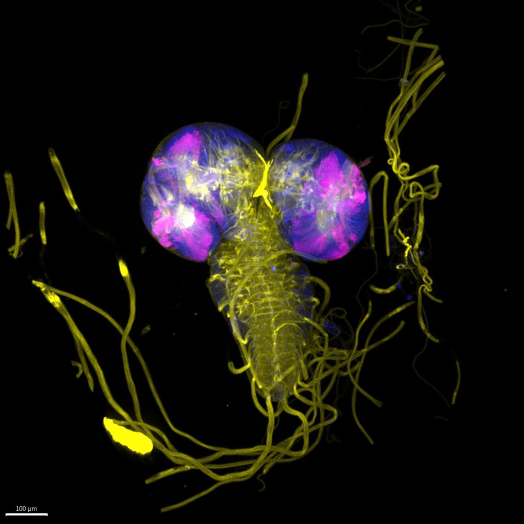
Our featured image, acquired by Maik Bischoff, captures a portion of the testis from the fruit fly Drosophila hydei. Like all insect testes, it is enveloped by a muscle layer. F-actin was stained using phalloidin, making the muscle fibers visible, while DAPI staining revealed the multiple nuclei within each syncytial myotube. An antibody against Drosophila

Featured image with Saurabh Chand Sagar

Posted by , on 25 April 2025

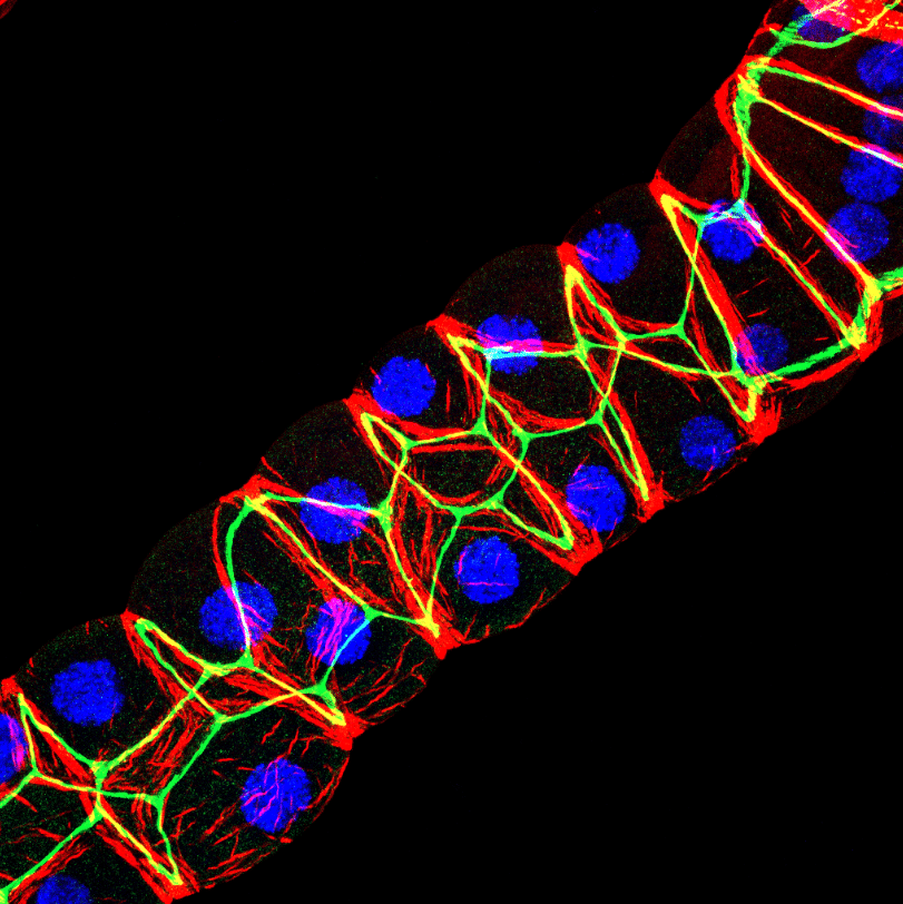
Our featured image, acquired by Saurabh Chand Sagar, presents a laser scanning confocal projection of the Malpighian tubules (MTs) from third instar wandering-stage (118 ± 2 hours post-eclosion) wild-type Drosophila melanogaster larvae. In the image, red denotes F-actin protein, green highlights Discs Large, a protein involved in maintenance of apicobasal polarity, and blue marks the

Inputs and Outputs of vEM in a Sensory System

Posted by , on 19 May 2023

by Federica Mangione, The Francis Crick Institute, UK DOI: 10.1242/focalplane.14809 Challenge The sense of touch enables organisms to interact with their environment by perceiving physical forces and guiding complex behaviors. Touch sensing is mediated by sensory neurons that innervate the body surface of animals and, together with surrounding cells, form specialized structures known as tactile

Featured image with Ellen Skarpen and José Teles Reis

Posted by , on 5 April 2023

Our featured image shows a Drosophila third instar larval brain with activated ‘Mosaic analysis with a repressible cell marker’ (MARCM) specifically in the Optic lobe, marked by GFP. This genetic technique allows for the labelling and manipulation of mitotically active clones of cells for the study of fate mapping or gene role through gain/loss of function. In this

Imaging nuclear dynamics and chromatin organization in a live intact organism: the design of the minimal constraint device

Posted by , on 9 March 2022

To visualize the response of the nucleus and chromatin to mechanical signals, I developed a device that enables their imaging in a live intact Drosophila larva.   Together with genetically encoded fluorescent markers, we can track muscle contraction and nuclear dynamics, as shown in Movie 1.  While the muscle is at rest, we can image chromatin

Development presents... an Imaging Special

Posted by , on 12 October 2021

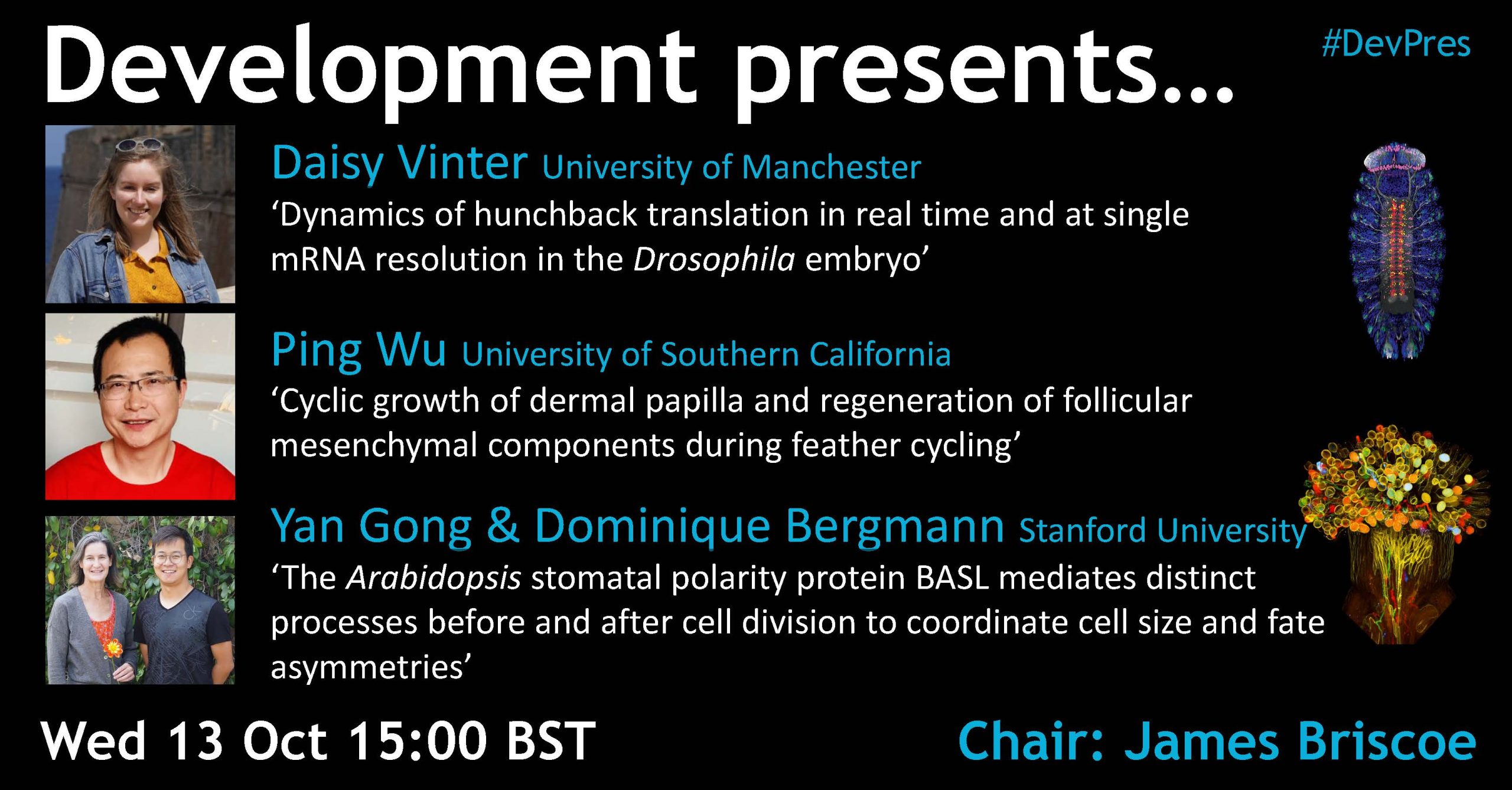
Development presents… is a webinar series hosted by Development. The webinars are chaired each month by a different Development Editor, who invites talks from authors of exciting new papers and preprints. First authors are particularly encouraged to present their work – we hope the series will become a forum for supporting early-career researchers. As well

Research Assistant Position

Posted by , on 26 June 2021

The Carver College of Medicine Department of Anatomy and Cell Biology is seeking a Research Assistant to join the Tootle lab (www.tootlelab.com) to perform research using Drosophila melanogaster (fruit flies) to define how lipid signals regulate actin in the cytoplasm and the nucleus. Execute planned research activities using observation, measurement and analytical techniques. Manage and maintain Drosophila