Advertisement

Displaying posts with the tag: is_archive

The Crick BioImage Analysis Symposium (2024): conference overview

Posted by , on 18 December 2024

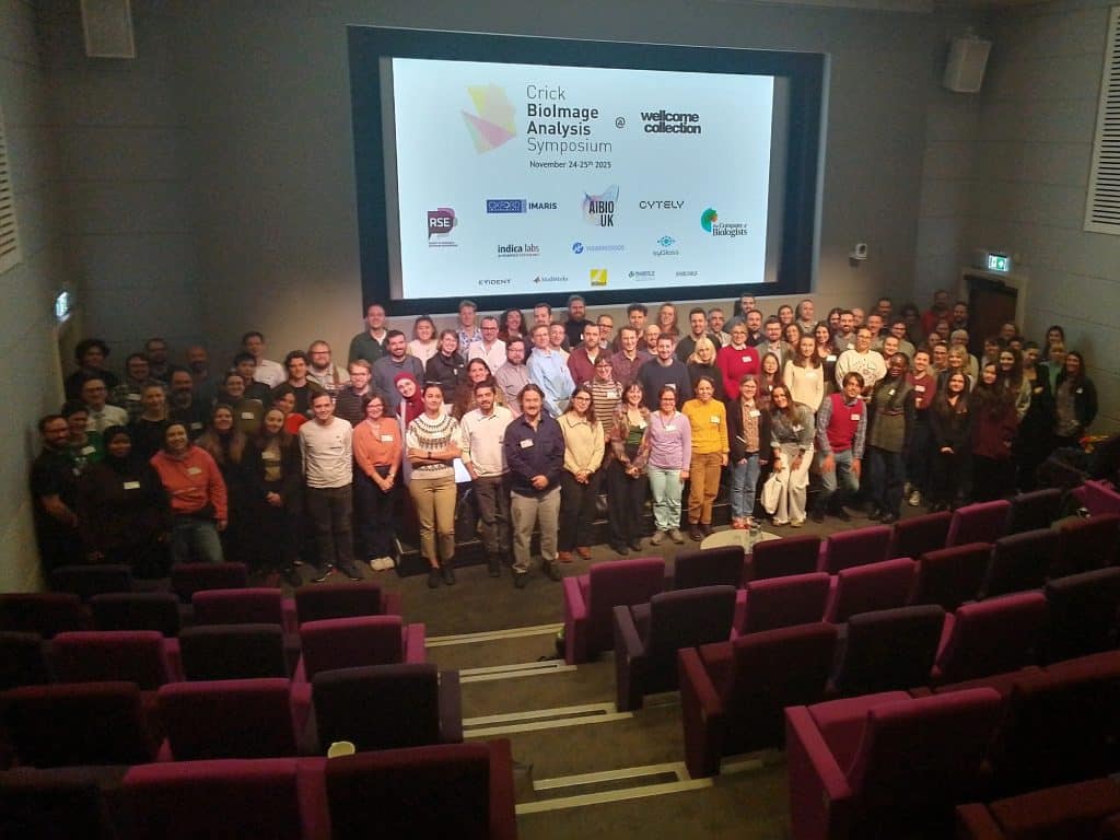
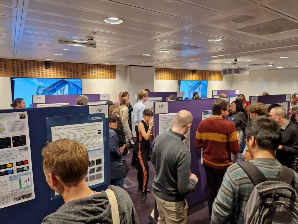
Have you missed this year’s Crick BioImage Analysis Symposium (CBIAS2024), or you want to know how this conference went to apply for next year’s edition? Here is our recap of this conference featuring impressions from 3 microscopists coming from different research backgrounds:   Zeinab Rekad, postdoctoral Research Associate, biologist, super-user and here for the 3rd time

Crick Bioimage Analysis Symposium 2023 - a Review

Posted by , on 22 December 2023

(By Vanessa Dao, Hradini Konthalapalli, Olatz Niembro Vivanco, Karishma Valand) The Crick Bioimage Analysis Symposium had its first in-person meeting in 2022. This year, #CBIAS2023 gathered around 200 people on site and 80 virtually. It has been an exciting two days of bringing biomedical researchers and their questions together with image analysis and their techniques.

Inputs and Outputs of vEM in a Sensory System

Posted by , on 19 May 2023

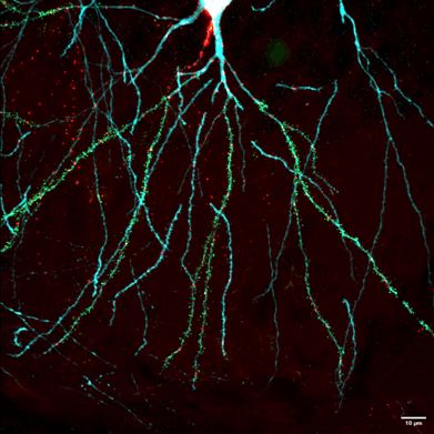
by Federica Mangione, The Francis Crick Institute, UK DOI: 10.1242/focalplane.14809 Challenge The sense of touch enables organisms to interact with their environment by perceiving physical forces and guiding complex behaviors. Touch sensing is mediated by sensory neurons that innervate the body surface of animals and, together with surrounding cells, form specialized structures known as tactile

Euro-BioImaging User Forum: Cardiovascular Research

Posted by , on 13 March 2023

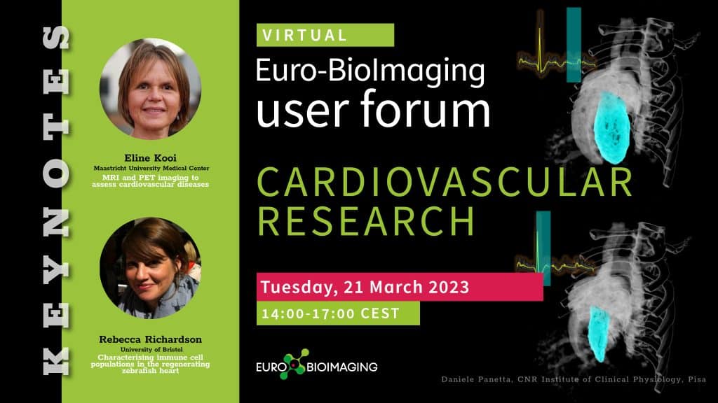
Euro-BioImaging is organizing a fifth online User Forum on Tuesday, March 21, 2023, from 14:00-17:00 CET. The topic is “Cardiovascular research.” This event will highlight the importance of cutting-edge imaging technologies in support of cardiovascular health, disease, diagnostics and the development of therapies. We will showcase the specific expertise available at our Nodes across Europe

AI4Life First Open Call

Posted by , on 20 February 2023

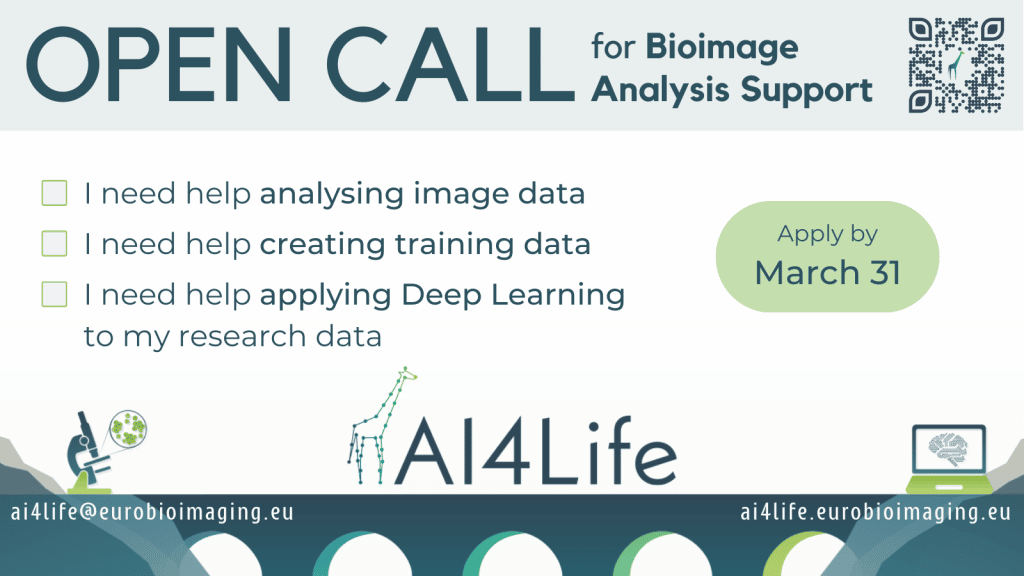
The European project AI4Life aims at narrowing the gap between life scientists performing biological imaging and developers of AI-based methods to analyze microscopy image data.  This is the first of a series of annual open calls, meant to provide life scientists who have unmet image analysis needs with adequate deep learning enhanced workflows for their

Crick BioImage Analysis Symposium (CBIAS) 2022 - a review

Posted by , on 14 December 2022

Did you want to participate in the Crick BioImage Analysis Symposium (CBIAS) but missed it? Are you planning to join us next year for CBIAS2023 but unsure if it would be relevant for you? We’ve asked three of this year’s participants to tell us about their experience and collated their reports here! We’ll hear from

Imaging Specialist - University of Warwick

Posted by , on 28 June 2022

Permanent Position, Full time – 36.5 hours per week The postholder will provide expert-level hands-on technical and user support for optical microscopy. The appointee will need to combine enthusiasm and careful attention to detail with adaptability. The post would suit an individual with a background in physics who wants to apply their skills to advance

Imaging Specialist, Columbia University, New York, NY

Posted by , on 3 December 2021

The Confocal and Specialized Microscopy Shared Resource at Columbia University Irving Medical Center is searching for an Imaging Specialist at the rank of Associate Research Scientist. This is a rare opportunity to join a highly rated, well-funded Comprehensive Cancer Center core that has provided advanced optical imaging expertise to hundreds of research labs for nearly

Postdoctoral Research Associate, Imaging Network, University of Münster (Germany)

Posted by , on 5 May 2021

The Microscopy Unit of the Imaging Network at the University of Münster, Germany, is seeking to fill the position of a Postdoctoral Research AssociateWissenschaftliche/r Mitarbeiter/in(salary level TV-L E 13, 100%) from 1 June 2021. We are offering a fixed-term full-time position for 5 years. Full-time employees are required to teach 4 hours per week during

LSFM series – Surfing on the data freak wave! Part II: Before imaging: Know your sample (geometry)

Posted by , on 10 October 2020

This post, part of the blog series "LSFM series – Surfing on the data freak wave!", discusses (a) Sample preparation, (b) Light interaction with matter, (c) sample alignment and (d) checking fluorescence and calibration