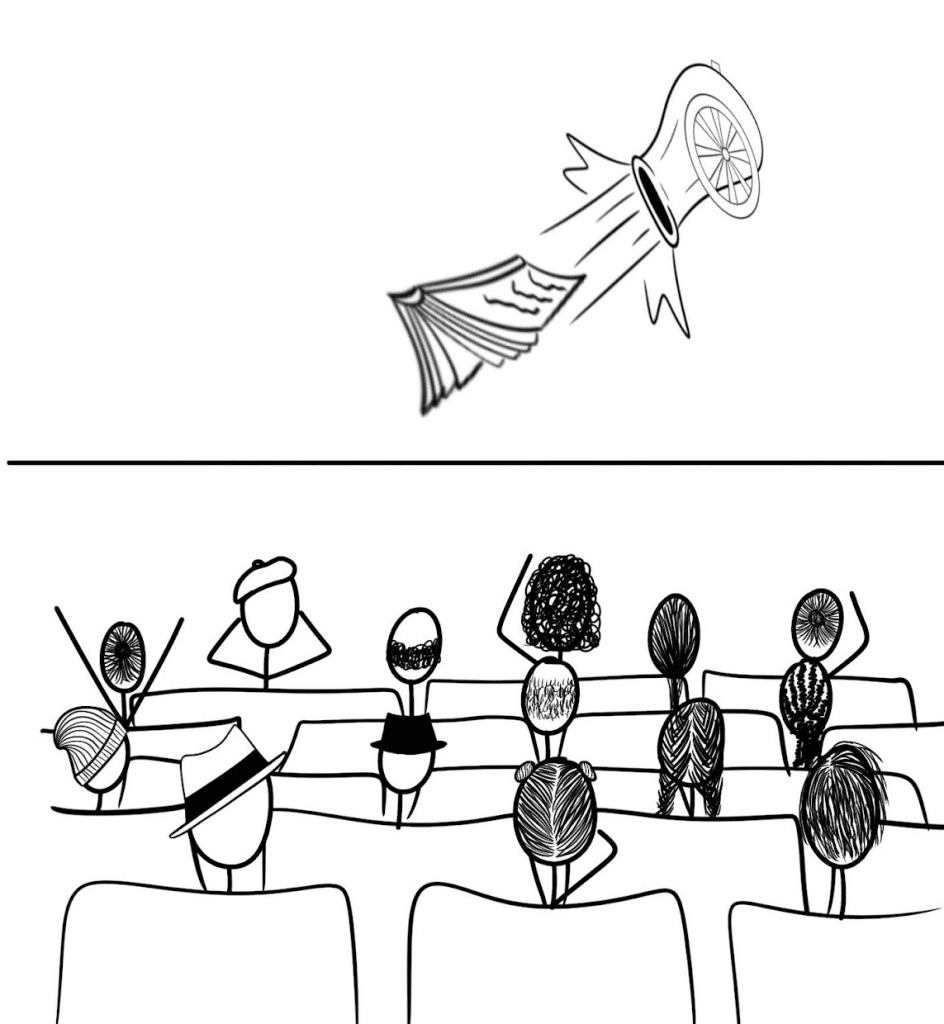
Crick BioImage Analysis Symposium (CBIAS) 2022 – a review
Posted by Stefania Marcotti, on 14 December 2022
Did you want to participate in the Crick BioImage Analysis Symposium (CBIAS) but missed it? Are you planning to join us next year for CBIAS2023 but unsure if it would be relevant for you? We’ve asked three of this year’s participants to tell us about their experience and collated their reports here! We’ll hear from Stefania, a postdoc and bioimage analyst in London, Zeinab, a PhD student in France, and Pradeep, bioimage analyst in Australia. They also chaired panels, presented posters and gave talks at the meeting, so really we are covering all bases!
Stefania Marcotti
It’s been a very exciting two days at the Crick Bioimage Analysis Symposium (#CBIAS2022, @The_CBIAS), hosted at the Francis Crick Institute in London. After two occurrences of the meeting in an online format, this year the hybrid format allowed some of us to meet in person for the first time after the pandemic. Needless to say, this made for an exciting opportunity for networking and for hearing about the latest development in the field of bioimage analysis. It was great to see more than 200 people from all over the world joining in person, plus about 70 online!
The varied program showcased invited speakers with expertise ranging from different imaging modalities to different computational methodologies, as well as shorter talks from more junior researchers. Questions from the audience after each talk received votes via Slack by both online and in-person attendees, and for each session the person asking the most upvoted question won a copy of the new open-access book “Bioimage Data Analysis Workflows” edited by Kota Miura. Companies also offered short pitches on their latest software developments, and it was very interesting to see some effort towards open science also from their side. The poster session over drinks and nibbles made for a great networking opportunity: topics covering anything from computational tool development, to user perspectives and infrastructure were discussed in what felt like a rather relaxed and welcoming atmosphere.
A highlight for me was Kota Miura’s talk titled “In Defence of the Integrity of Bioimage Analysis”, where he outlined best practices, common problems with deliberate and inadvertent data fabrication, and mistakes that might lead to mistrust towards the work of bioimage analysts. The discussion of this talk and some of the others revolved around the need for good documentation of computational tools, and precise writing up of method sections in papers where these tools are used. Only a joint effort between developers, analysts and users can produce better science!
But what is a bioimage analyst? The first panel of the meeting, chaired by Siân Culley (King’s College London), saw Colby Benari (In2Science), Dale Moulding (University College London) and Helen Zenner (FocalPlane) attempting to find a definition to the role of bioimage analyst. Interestingly, Colby pointed out that young students looking at college and university options, often struggle to see the variety of roles that STEM has to offer, and tend to look at other disciplines if being a medical doctor or a wet lab scientist does not appeal to them. Other topics were discussed more centred around the role of bioimage analysts as a bridge between experimental scientists and computational developers: it was agreed that a one-size-fit-all definition might be difficult to find, and that often the job title encompasses a range of roles, skillsets, and degrees of involvement with the scientific questions.
So, should you aim to become a bioimage analyst? I had the honour of chairing the panel at the end of the second day, with Robert Haase (TU Dresden), Arianne Bercowsky (DotPhoton) and Kota Miura (University of Heidelberg) evaluating this question. The answer, after a very lively discussion among the panellists and the audience, turned out to be similar to the answer to other very important questions: it depends! Ultimately on your interests, and on the direction you’d like your career to go. A point made by Arianne really stuck with me: when asked whether the role of bioimage analysts is actually needed, she questioned the difference between using Google translate versus knowing the spoken language when visiting a foreign country. While simple communication would probably be successful in both cases, more complex interactions would only be allowed when a common language is spoken. Similarly, the bioimage analyst can ensure effective communication between experimentalists and developers, and increase the depth of successful use of scientific data.
So, to sum up, we might not all agree on the definition of a bioimage analyst and if you ask us if you should become one, we might answer that it depends, but the sense of community is strong, and this is what is making a difference! Looking forward to CBIAS2023 already!

Zeinab Rekad
#CBIAS2022 from the lens of a biologist:
I’m a Ph.D. wet lab scientist working on cell adhesion and extracellular matrix remodeling. I started doing image analysis about two years ago when it became apparent that my project would heavily rely on multiple imaging modalities and techniques. This year’s first in-person symposium was also my first time attending the symposium.
I first would like to highlight how this #CBIAS2022 edition was very well-organized: session length, breaks, networking time etc., all were on point. I also enormously appreciated the smooth inclusion of sponsors throughout the conference by alternating between plenary sessions and “tech bites” and setting up a sponsor quiz. To end on the organization aspect, and as a young scientist trying to fight growing ecological challenges, I hugely applaud the organizer’s effort to reduce unnecessary waste during the conference. Washable glass cups, metal cutlery, and ceramic plates were much appreciated and a good change from the “traditional” disposable plastic tableware.
On the scientific side, and as a wet lab scientist, I have long questioned my participation in this conference as I was afraid of not having enough data to share nor being “an expert” in developing any image analysis pipeline/tool.
However, biologists often forget that many of today’s advances in bio-image analysis are driven by the evolution and complexity of our biological questions. Thus, a biologist able to understand key concepts and challenges of this young field is (for now) quite rare and appreciated within the community.
Indeed, I felt more than welcome and enjoyed the conference as much as the ‘developers’. This symposium was, for instance, a golden opportunity for biologists like me to participate in discussions with the community, share my needs, and give feedback as a user of multiple image analysis tools. Having the opportunity to extend my small network was also a huge advantage, as I tend to have less in-person interaction with the bio-image analysis community at my home institution. I also enjoyed the poster session’s friendly and sometimes geeky atmosphere, which was a great opportunity to learn about the new methods and tools that could help me extract biologically relevant information from my images.
From this conference, I also got an overview of what are today’s challenges in the bioimage analysis field as the complex manipulation of 3D images (Robert Haase)/ Applying the FAIR principles to AI-mediated image analysis (Estibaliz Gómez de Mariscal)/ as well as our collective responsibility in the exploitation of our images (Kota Miura). As Stefania Marcotti nicely highlighted, I also particularly enjoyed the panel discussions. Indeed, hearing the perspectives of the guests and attendees made me realize how much the bio-image analysis field is driven by researchers coming from multiple backgrounds and with diverse expertise. And I can only be optimistic for the future as seeing such scientific exchange made me reflect on how billion years ago, life thrived thanks to cooperation and symbiosis between all kinds of life forms.
Although I do not recommend attending the symposium for the absolute beginners in image analysis (to the risk of feeling overwhelmed and a bit lost), I had the most fantastic and enjoyable time a scientist can have at a conference. Thus, biologists with a good foundation in image analysis principles (image filtering, general view of segmentation methods, and basic knowledge of main image analysis tools, which all can be learned from Neubias network seminars), attending the Crick Bio-Image Analysis Symposium is the step you should take to move forward in your image analysis journey.
Pradeep Rajasekhar
I had the privilege of attending and presenting at the Crick Bioimage Analysis Symposium (CBIAS) 2022, at the Francis Crick Institute in London. It was a super long trip for me as I’m based in Melbourne, Australia at the Walter and Eliza Hall Institute of Medical Research. As there is a strong bioimage analyst presence in UK and Europe, it was a great chance to network, gain exposure and importantly, meet the famous bioimage analysts I’m so familiar with from Twitter everyday, in 3D!
I attended NEUBIAS 2020 as a postdoctoral scientist who was doing wet lab work, microscopy and image analysis. I was at a crucial juncture in my career, as it inspired me to specialize in bioimage analysis. I must admit that meeting Robert Haase at NEUBIAS and becoming collaborators played a major part. CBIAS was the first major conference I attended as a bioimage analyst and where I presented one of my first bioimage analysis software projects. With support from the Chan Zuckerberg initiative, we developed a napari plugin for processing lattice lightsheet data, napari-lattice. I was nervous to be presenting after Robert Haase, but it was the perfect slot as our plugin uses the tools he has developed. I got some great feedback, especially during the networking sessions, and identified some opportunities for growth. One thing I learnt (again!), always have a backup of your presentation on a USB drive.
CBIAS had a great mix of bioimage analysis talks from various fields, such as studying ear morphogenesis in zebrafish to quantifying DNA structure using Atomic Force Microscopy (AFM). The breadth and depth of the analysis approaches employed, and the biological insights generated were fascinating. As expected, there were a lot of talks on deep-learning, but a major highlight was DeepImageJ and the Bioimage Model Zoo by Estibaliz Gómez de Mariscal. She talked about the importance of bridging the gap between those who develop specialized deep-learning models and the end-users who apply it in specific biological contexts, especially within the Fiji ecosystem.
The panel discussions on both days were informative and on the second day, Stefania Marcotti who did an amazing job as a moderator brought up a question of whether you should learn programming to be a bioimage analyst. I think this was a highly divisive topic, which in turn makes it fun! It generated a lot of discussion, as there were strong arguments for and against, and at the end we “mostly” settled on “It Depends”. This in itself highlights the diversity of the bioimage analysis field in terms of people, biological problems to be solved and the software tools available. I think for most research questions, you can use software like Fiji and Imaris without touching any code. However, if you’ve got the time and patience, learning coding unlocks a whole new world of technical and career possibilities.
Although not a part of the conference, another highlight of my trip was being a part of a hackathon organized by Robert Haase’s team from TU Dresden and Jonathan Smith at the Crick Institute. I learned a lot interacting with seasoned developers, understanding their thought process and how they approach problems. We worked on developing tools within the clesperanto ecosystem, such as implementing user-friendly ways for GPU-accelerated image processing on high performance computing systems (HPC), how to program in OpenCL and ways to contribute to pyclesperanto. During the week, Martin Jones and Rocco D’Antuono from the Crick Institute were kind enough to give us a tour of the imaging facility and data centre.
Overall, it was a fantastic learning experience for me and I would like to thank the organizers of the Crick Bioimage Analysis symposium for doing a great job. I think this would be a great conference for biologists who use microscopy and seasoned bioimage analysts alike. I do admit it can get quite technical and overwhelming for a biologist, but meetings like these give you an opportunity to network with bioimage analysts, and see what tools and approaches are out there. It is likely that an analytical approach from a different field would be a good fit. All it takes is one good collaboration to make a difference!


 (9 votes, average: 1.00 out of 1)