Advertisement

Displaying posts with the tag: is_archive

Featured image with Theresa Wiesner

Posted by , on 21 June 2024

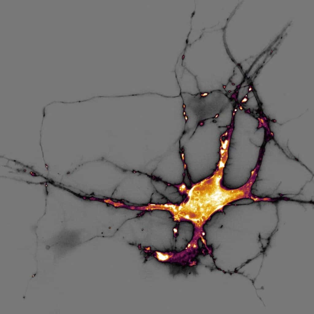
Our featured image, acquired by Theresa Wiesner, shows a neuron in dissociated hippocampal culture and was imaged using highly inclined thin illumination microscopy (HiLO). The neuron is transfected with a neurotransmitter (glutamate) sensor and shown in orange. The image was acquired using Nikon Elements software / microscope and post-processing was done using FIJI (ImageJ). Find

Journal of Cell Science Special Issue: Cell and Tissue Polarity

Posted by , on 18 March 2024

The Journal of Cell Science Special Issue on Cell and Tissue Polarity is now complete. The original research in this Issue, guest edited by David Bryant, covers many aspects of cell polarity, including the roles of both the cytoskeleton and junctions in polarity establishment and maintenance, polarised membrane trafficking, cell division and collective cell migration.

A general strategy to develop fluorogenic polymethine dyes for bioimaging

Posted by , on 6 December 2023

by Annabell Martin and Pablo Rivera-Fuentes. Fluorescence imaging is an invaluable tool to study biological processes and further progress depends on the development of novel advanced fluorescent probes. Excitingly, using polymethines as a scaffold for fluorogenic dyes, we have opened up a lot of new avenues for bioimaging.

Featured image with David McGrath

Posted by , on 21 July 2023

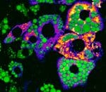
Our featured image is of mature mouse brown adipocytes (WT-1 cell line) under normal conditions from David McGrath. Channel 1 (green LUT) shows lipid droplets stained with BODIPY 493/503 and channel 2 (fire LUT) shows mitochondria stained with TMRE. The image was obtained using a Visitron SD-TIRF confocal microscope (60x Apo TIRF (corr.) Oil/ NA: 1.49) at the UKE Microscopy Imaging Facility

Imaging Specialist - University of Warwick

Posted by , on 28 June 2022

Permanent Position, Full time – 36.5 hours per week The postholder will provide expert-level hands-on technical and user support for optical microscopy. The appointee will need to combine enthusiasm and careful attention to detail with adaptability. The post would suit an individual with a background in physics who wants to apply their skills to advance

Phototoxicity - the good, the bad and the quantified.

Posted by , on 14 May 2021

Our virtual meeting on phototoxicity was held in late January 2021, generously sponsored by the European Microscopy Society and enabled by the Royal Microscopical Society. In four hours, spread over two days, the five organisers and twenty invited participants discussed the problem of phototoxicity in live imaging, and how we can start to tackle this

Imaging Specialist - University of Warwick, UK

Posted by , on 22 February 2021

Fixed Term Contract until 30 September 2022 The postholder, funded through a Wellcome Multi-user equipment award, will provide expert-level hands-on technical and user support for lattice light sheet microscopy. The appointee will need to combine enthusiasm and careful attention to detail with adaptability. The post would suit an individual with either a background in physics

Technology highlights - Quantitative Phase Imaging (QPI)

Posted by , on 20 January 2021

Interview with Helena Chmelová, Ph.D. from the Light Microscopy Core facility at the Institute of Molecular Genetics of the Czech Academy of Sciences in Prague, Czech Republic. Can you tell us a little bit about yourself – where you work, what your research focus is? I work at the Light Microscopy Core Facility at the Institute of