Featured image with David McGrath
Posted by FocalPlane, on 21 July 2023
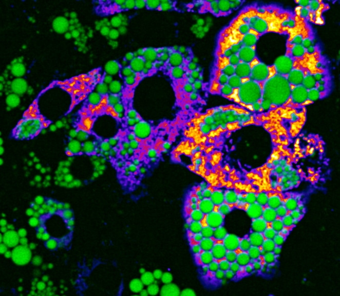
Our featured image is of mature mouse brown adipocytes (WT-1 cell line) under normal conditions. Channel 1 (green LUT) shows lipid droplets stained with BODIPY 493/503 and channel 2 (fire LUT) shows mitochondria stained with TMRE. The image was obtained using a Visitron SD-TIRF confocal microscope (60x Apo TIRF (corr.) Oil/ NA: 1.49) at the UKE Microscopy Imaging Facility (UMIF) in Hamburg, Germany. Image processing and adjustment were done using Fiji ImageJ.
Author Information: David McGrath, PhD candidate at the CellComM and Heeren labs, Institute of Biochemistry and Molecular Cell Biology, UKE, Hamburg, Germany
@DavidMcGrath_9, @cellcommlab @pj_saez, @JoergHeeren, @Endo_Connect
Authors acknowledge and thank A. V. Failla and the UKE Microscopy Imaging Facility (UMIF) under the DFG Research Infrastructure Portal: RI_00489.
We caught up with David to find out more about his research career and what he is excited about in microscopy.
Research career so far: My research career started with my BSc in Sports and Exercise Science, at the University of Limerick in Ireland, where I developed an interest in the molecular mechanisms by which different forms of exercise can remodel skeletal muscle tissue to benefit metabolic health. This interest lead me to study Biomedical Sciences at Maastricht University, The Netherlands, where I completed my master’s degree. Here, my research focused on various aspects of metabolic health, such as the role of mitophagy on mitochondrial function in iron deficient skeletal muscle cells, and understanding the role of myocardial energetics in the development of diabetic cardiomyopathy.
Current research: Currently, I am doing my PhD in the labs of Professor Pablo J. Sáez (CellComM lab; @cellcommlab, @pj_saez) and Professor Jörg Heeren (@JoergHeeren). I am investigating the role of endo-lysosomal membrane proteins for energy metabolism in white and brown adipose tissues. My project mainly utilises optical microscopy techniques and advanced imaging analysis to determine features such as lipid trafficking and organelle dynamics. This project is part of an international training network consortium, called EndoConnect (@Endo_Connect), which is funded from the European Union’s Horizon 2020 Research and Innovation programme under the Marie Skłodowska-Curie Grant Agreement No. 953489.
Favourite imaging technique/microscope: At present, my favourite imaging technique is live-cell imaging using spinning disk (Visitron SD-TIRF confocal microscope), which I use to visualise organelle dynamics and interactions. It is really enjoyable to open the timelapse file and to watch the multiple events occurring before my eyes.
What are you most excited about in microscopy?
I am excited about the development of live-cell super resolution microscopy and fluorescent multiplexing. In the future, I look forward to seeing how far deep learning image analysis software develops to help microscopists to understand complex dynamic processes.
 (No Ratings Yet)