Advertisement

Displaying posts with the tag: is_archive

Microscopy preprints – new tools and techniques in imaging

Posted by , on 2 May 2025

Here is a curated selection of preprints posted recently on new tools and techniques in imaging. Let us know if we are missing any recent preprints that are on your reading list!

Microscopy preprints – new tools and techniques in imaging

Posted by , on 9 August 2024

Here is a curated selection of preprints posted recently on new tools and techniques in imaging. Let us know if we are missing any recent preprints that are on your reading list!

Affimers – small probes for big imaging experiments

Posted by , on 9 August 2022

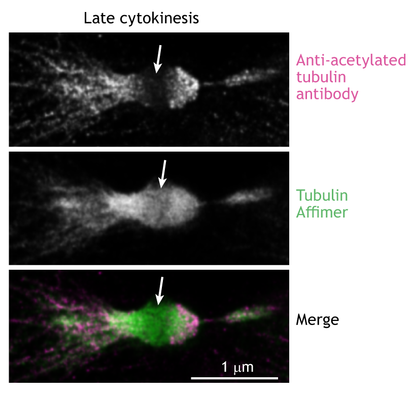
Antibodies have been used to label proteins of interest since the 1970s. However, their large size (around 15 nm long for IgG) makes the penetration of dense structures within the cell a challenge and places the fluorophore away from the target protein. This latter issue, known as a ‘linkage error’, particularly impacts accuracy and precision

A new Review in Journal of Cell Science discusses how specimens can impact image acquisition

Posted by , on 13 April 2022

With the development of new imaging strategies and technologies and advanced computational analyses has come a body of literature addressing various aspects of optimising imaging and processing. However, the impact of the sample itself on the quality of the image attained has garnered less exploration. Fluorescent microscopy is dependant on two factors – the intensity

Imaging nuclear dynamics and chromatin organization in a live intact organism: the design of the minimal constraint device

Posted by , on 9 March 2022

To visualize the response of the nucleus and chromatin to mechanical signals, I developed a device that enables their imaging in a live intact Drosophila larva.   Together with genetically encoded fluorescent markers, we can track muscle contraction and nuclear dynamics, as shown in Movie 1.  While the muscle is at rest, we can image chromatin

Protein micropatterning: beauty standards in cell culture

Posted by , on 16 February 2022

When imaging cells grown on a flat substrate, such as a glass coverslip, we quickly admire the diversity of morphologies different cells can take on. Some cells, such as COS7, will take on a flat “pancake”-like shape, which makes it easy to image its organellar structure in a 2D plane. On the other hand, a

Asymmetric cell divisions in 3 dimension stem cell colonies

Posted by , on 27 September 2021

When looking at the development of a multicellular organism, for example a human, the first striking feature is the progressive increase in cell numbers due to successive divisions, from one single tiny cell 80 mm in diameter to a 3.5 kg ball of organised cells forming a new-born baby. The way cells divide, in particular

Using Nile Blue and Nile Red to visualise the presence of lipids in cryosectioned tissues

Posted by , on 14 September 2021

Lipids are crucial elements of mammalian (and non-mammalian) cell biology and yet lipids are challenging to visualise in situ. In comparison to proteins, which we can generate antibodies for, or carbohydrates, some of which we can detect using fluorescent lectins, there are relatively few lipid-specific fluorescent probes. Many lipids are highly conserved across species making

Tissue clearing: what invisible samples reveal about biology

Posted by , on 8 September 2021

Written by Jorge Almagro and Hendrik Messal The tissues that constitute organs exist in our body in 3D. However, for practical reasons, histological analysis has been traditionally performed in 2D, slicing a few micrometer-thick sections to analyse them under the microscope. While this has been critical to understand our anatomy for centuries, it constitutes a simplification of

What is Expansion Microscopy?

Sponsored by Andor, on 27 May 2021

How can you get the most information from expanded samples? Traditional light microscopy is limited by the diffraction of light, consequently, features less than 200 nm apart cannot be resolved. For a significant time microscopy technique development was focused towards improving imaging techniques to allow for individual molecules to be resolved. Super-resolution microscopy was developed