Advertisement

Displaying posts with the tag: is_archive

Microscopy preprints - bioimage analysis

Posted by , on 30 May 2025

Here is a curated selection of preprints published recently (up until 22 May). In this post, we focus specifically on bioimage analysis and data management.

VolumeEM: An Interview with Kedar Narayan

Posted by , on 14 December 2024

I had the pleasure of sitting down with recent awardee of the Alan Agar Royal Microscopy Award for Electron Microscopy, Dr. Kedar Narayan. Dr. Narayan is very involved with the grassroots Volume EM (vEM) community, and has used his time at Frederick National Laboratory to not only benefit cancer research, but microscopy technique as a

Featured image with Jiwon Lee

Posted by , on 6 December 2024

Our featured image, acquired by Jiwon Lee, is a 3D rendering model of Plasmodium falciparum showing the complex organisation of cellular organelles within the malaria parasite. Using a focused ion beam scanning electron microscope (FIB-SEM), a series of high resolution 2D images were acquired. The image stack was aligned and each organelle was segmented in

Microscopy preprints - bioimage analysis

Posted by , on 29 November 2024

Here is a curated selection of preprints published recently. In this post, we focus specifically on bioimage analysis and data management.

FocalPlane features... Imaging Cell Architecture and Dynamics with Journal of Cell Science

Posted by , on 15 November 2024

Our latest FocalPlane features… webinar was a celebration of the research in Journal of Cell Science’s Special Issue: Imaging Cell Architecture and Dynamics. Our Guest Editors Lucy Collinson and Guillaume Jacquemet hosted the webinar with talks from Md Hashim Reza, Melanie Rug and Vikas Tillu.

Journal of Cell Science Special Issue: Imaging Cell Architecture and Dynamics

Posted by , on 5 November 2024

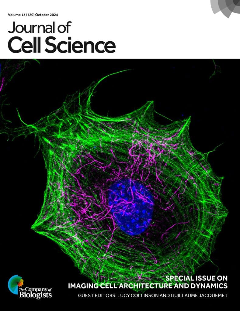
Journal of Cell Science’s Special Issue on Imaging Cell Architecture and Dynamics is now complete. The original research in this Issue, guest edited by Lucy Collinson and Guillaume Jacquemet, covers a diverse range of imaging methodologies including CLEM, super-resolution imaging, volume EM, high-throughput imaging, FRET, expansion microscopy, mesoscopy and immuno-SEM, as well as showcasing new

vEM reveals the cellular composition of the Blood Nerve Barrier

Posted by , on 11 October 2024

Using Serial Block-Face-SEM6 and AT-SEM, we characterized the cellular composition and interactions along EndoBVs7. Unlike the BBB, we observed that the vasculature within the endoneurium is not completely covered. Upon closer examination of the cells in contact with EndoBVs, we identified three distinct cell types frequently interacting with the vasculature.

Kidney elastic fiber network changes in Marfan's Syndrome

Posted by , on 19 July 2024

by de Souza RB*, Meek KM#, Lewis PN#, Pereira LV*. *Department of Genetics and Evolutionary Biology, University of São Paulo, Brazil #StructuralbBiophysics Research Group, School of Optometry and Vision Sciences of the United Kingdom, Cardiff University, UK Challenge Marfan’s syndrome (MFS) is an autosomal dominant disorder that affects the connective tissue, resulting from a mutation

Targeted vCLEM of heterogeneous samples

Posted by , on 15 January 2024

by Karel Mocaer* and Paolo Ronchi# *Schwab Team and #EMCF, EMBL Heidelberg, Germany Challenge Studying cells or events within their biological context (i.e. in tissues, ecosystem etc…) is highly relevant. However, volume EM techniques are limited in the volume that can be acquired. Therefore, targeting approaches are needed to characterize specific regions of interest. This

Deciphering cardiac multiscale organization

Posted by , on 11 December 2023

by Sandra Rugonyi and Claudia López Oregon Health & Science University, Portland Oregon, USA Challenge The morphology, microstructure, and ultrastructure of heart tissues is exquisitely organized to optimize cardiac efficiency. In cases of cardiac malformations, known as congenital heart disease (CHD), spatially heterogeneous and abnormal ultrastructural patterns emerge as early as fetal stages that worsen