Advertisement

Displaying posts with the tag: is_archive

Featured image with Shivangi Verma

Posted by , on 30 August 2024

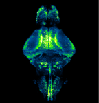
Our featured image, acquired by Shivangi Verma, gives us a peek into the brain of a zebrafish. It depicts neurons in the whole brain of a larva expressing GCaMP6f, a fluorescent reporter of neuronal activity, using a pan-neuronal promoter. A 10-day old transparent homozygous albino larva was embedded in agarose and imaged using ZEISS Lightsheet

Featured image with Upasana Gupta

Posted by , on 2 August 2024

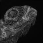
Our featured image, acquired by Upasana Gupta, represents the cellular architecture of zebrafish (larva) eye. The tissue was prepared by paraformaldehyde fixation of a 4-day post fertilisation larva, followed by staining with Hoechst nuclear stain. The sample was imaged and processed using Leica SP8 confocal microscope and Leica software at the Bio-imaging facility at the

See more with HCR spectral imaging

Posted by , on 6 March 2024

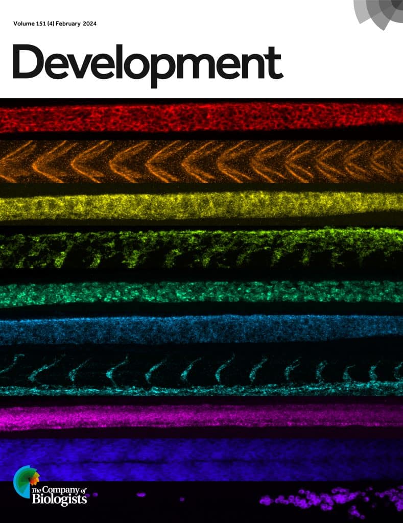
In a new paper published in Development, Samuel Schulte, Niles Pierce and colleagues present a new workflow for HCR spectral imaging with linear unmixing, for simultaneous imaging of ten RNA and/or protein targets in both whole-mount zebrafish embryos and mouse brain sections.

Bioimaging Neurofilaments in vivo (Postdoc or Engineer)

Posted by , on 18 June 2021

In the context of an ERC funded project, the laboratory of Dr. Bomont (NeuroMyoGene institute, Lyon, France) is recruiting several fellows (2 postdoc, 1 technician/engineer). In particular, we are seeking highly motivated candidate who wish to apply advanced live microscopy methodologies to study the dynamics of neurofilaments in vivo. This project is multidisciplinary and will be

Postdoctoral Fellow position in Bioimaging Neurofilaments in vivo

Posted by , on 18 February 2021

In the context of an ERC funded project, the laboratory of Dr. Bomont (NeuroMyoGene institute, Lyon, France) is recruiting several fellows. In particular, we are seeking highly motivated candidate who wish to apply advanced live microscopy methodologies to study the dynamics of neurofilaments in vivo. This project is multidisciplinary and will be conducted in an