And the winner is…
Posted by FocalPlane, on 1 September 2023
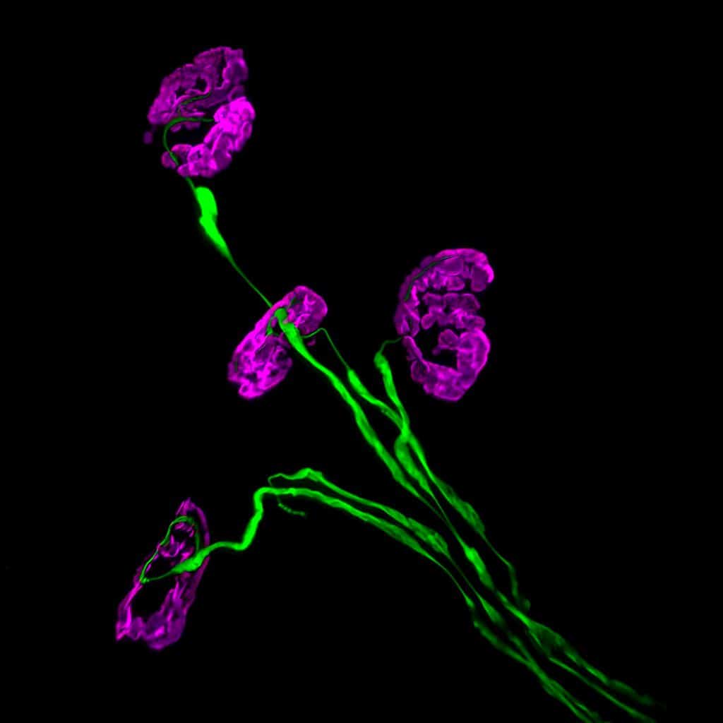
We are delighted to announce that the winner of the 2023 FocalPlane image competition is Rebecca Simkin. Rebecca’s image, ‘Neuromuscular junctions’, depicts four NMJs in a lumbrical muscle, located in the hind paw of a wildtype mouse. A wholemount immunostaining was performed using 2H3 and SV2 antibodies to visualise lower motor neurons in green (specifically neurofilament and synaptic vesicles, respectively), and α-bungarotoxin to label post-synaptic acetylcholine receptors in magenta. The image was captured on a LSM780 confocal microscope and is a z-projection of maximum intensity. Enhancement of colours was performed using Fiji, ImageJ. ‘Neuromuscular Junctions’ was acquired in the Department of Neuromuscular Diseases at the UCL Queen Square Institute of Neurology (QSIoN; @UCLIoN). The NMJ – a chemical synapse between a motor neuron and muscle fibre – is particularly vulnerable and undergoes degeneration as an early pathogenic event in amyotrophic lateral sclerosis (ALS), a devastating neurological disease in which progressive degeneration of motor neurons induces muscle paralysis, and eventually death.
Rebecca’s image will be appearing on an upcoming issue of Journal of Cell Science.
We caught up with Rebecca to find out more about her research career so far, her current research, what excites her in microscopy and her future plans.
What first inspired you to become a scientist?
It was my A-level chemistry teacher that primarily sparked my curiosity in science; he was incredibly enthusiastic and passionate about the subject, and made the practical element entertaining. I was always motivated by the challenge of needing to understand mathematical concepts and physical theories in chemistry, rather than simply memorising facts. This led me to pursue an undergraduate degree in pharmacology, where I was then introduced to laboratory research.

Can tell us about your research career so far?
I completed my BSc in Pharmacology at the University of Bristol in 2020, where I gained first-hand experience in a research laboratory. For my final year project, I helped to characterise the pharmacological profile of several drugs – elinogrel, AZD-1283 and PSB-0739 – at the P2Y12 receptor under the supervision of Professor Stuart Mundell. Following this, I moved back to London and joined Dr James Sleigh at the UCL QSIoN to obtain an MRes in Translational Neuroscience. My research involved characterising sensory neuron phenotypes of CRISPR/Cas9-generated mouse models of two diverse forms of Charcot-Marie-Tooth disease, a heterogeneous group of genetic peripheral neuropathies. I have now extended my studies to in vivo investigations of early ALS pathogenesis, another condition that affects the neuromuscular system.
What are your currently researching?
I began my PhD with Professor Pietro Fratta (@FrattaLab) in February 2022, and I am currently in my second year. My research aims to investigate how mutations in RNA-binding proteins lead to elevated spinal motor neuron (sMN) vulnerability in ALS. I hope to establish whether downregulation of the synaptic protein UNC13A – as detected in human TDP-ALS models – affects neuromuscular junction physiology and innervation, determine the transcriptomic alterations occurring in sMNs of a humanised FUS-ALS mouse model, and establish how this particular FUS-ALS mutation impacts the sMN response to stress (i.e., following nerve injury).
Where do you see yourself in five years?
I hope to have obtained my PhD, and be a postdoctoral researcher.
What is your favourite imaging technique/microscope?
I’ve always used the LSM780 or LSM980 confocal microscope for my experiments. Although I haven’t performed live-cell imaging myself, I have observed in vivo axonal transport of HcT-tagged signalling endosomes in the sciatic nerve. Being able to visualise biological processes in real-time at the molecular level is simply fascinating to me!
What are you most excited about in microscopy?
I am particularly excited about the integration of artificial intelligence (AI) with confocal microscopy, and single-cell and spatial transcriptomics. AI-driven advancements have enhanced the quality of captured images (i.e., through de-noising and optimising parameters), and accelerated data acquisition and interpretation. Other novel techniques, such as integrating MERFISH with confocal microscopy, has enabled researchers to study gene expression patterns within intact tissues at the single-cell level, and provides an insight into the spatial organisation of cells.
How did you hear about the FocalPlane image competition?
I initially heard about the FocalPlane image competition through my secondary supervisor, Dr Nicol Birsa. She forwarded FocalPlane’s tweet about the upcoming deadline, and I applied instantly!
Images such as yours are a fantastic way to engage people with science; do you have any plans to get involved in more sciart/scicomm in the future?
Although I have nothing lined up just yet, I would love to get involved in more SciArt and SciComm. I plan to continue entering image competitions such as these, and join some public outreach events, e.g., Soapbox Science. It would also be great to write scientific blog posts, or eventually dabble in scientific journalism!
Thanks to everyone who entered our image competition and congratulations to our winner, Rebecca Simkin. Her striking image of neuromuscular junctions will be the cover image on an upcoming issue of Journal of Cell Science. Also, look out for our fortnightly ‘Featured image‘ posts to find out more about some of the scientists that entered our competition.
 (No Ratings Yet)