Featured image with Oona Paavolainen
Posted by FocalPlane, on 15 November 2023
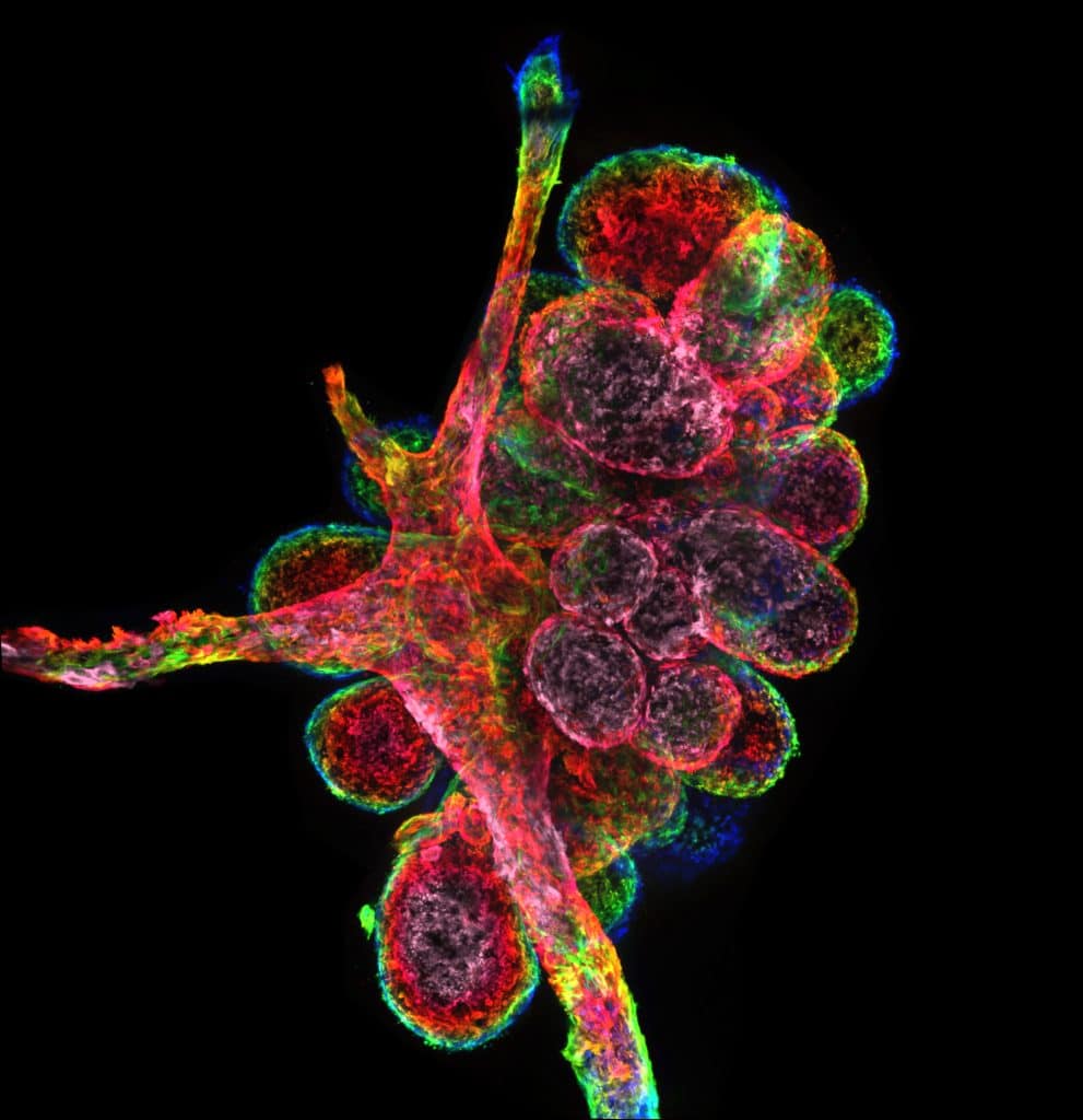
Our featured image, acquired by Oona Paavolainen, shows a primary mammary gland organoid, in a collagen-I matrix, formed from a single primary epithelial cell isolated from material obtained from a breast reduction surgery. The image was taken with a spinning disk confocal microscope (3i CSU-W1 Spinning disk) using a 20x Zeiss Plan-Apochromat (NA=0.8) objective. The constructed image was made in ImageJ and is a maximum intensity projection with a rainbow depth-coded lookup-table.

We caught up with Oona to find out about her research and what she is excited about in microscopy.
Research career so far: I’ve completed a bachelor’s in biomedical sciences in the UK, and a Masters in biomedical imaging in Finland. Currently, I am a 4th year PhD candidate at the University of Turku in Finland, in the laboratory of Dr Emilia Peuhu.
Current research: Our lab works on uncovering regulatory cues and mechanisms determining cell fate during postnatal development and malignant growth in the mammary gland. My PhD work, in particular, has focused on deciphering the 3-dimensional architecture of the terminal ductal-lobular units of the human mammary gland, and the factors that determine this shape during normal development. Breast tissue holds an extensive capacity for differentiation through the lifetime, and understanding its shape, the spectrum of cellular identities, and the dynamic environmental niches that contribute to it can shed light on malignant transformation as well.
Favourite imaging technique/microscope: I always say that a day I get to spend time on a microscope is a good day at work. In particular, I enjoy the challenge of figuring out which instrument provides me with the necessary information to answer a question. I work with a large range of samples and resolution requirements from whole tissue samples to intracellular components. I play no favourites – I’ve learned that any technique, if utilised well, can make me go ‘wow’! And that’s what keeps me coming back.
What are you most excited about in microscopy? I’m really excited about the possibilities in combining different techniques. Yielding information that is traceable to an expression or a spatial location in a sample can be very powerful, as demonstrated by spatial transcriptomics, especially in the context of heterogeneous samples such as tissues. As imaging techniques get less damaging, what else can we extract from the same sample? Much nuance is averaged out when working with singular techniques. Directly complimentary information could be very powerful and I think that the sky is the limit for our imagination when it comes to utilising samples to the fullest!


 (7 votes, average: 1.00 out of 1)