Featured image with Till Stephan
Posted by FocalPlane, on 14 November 2023
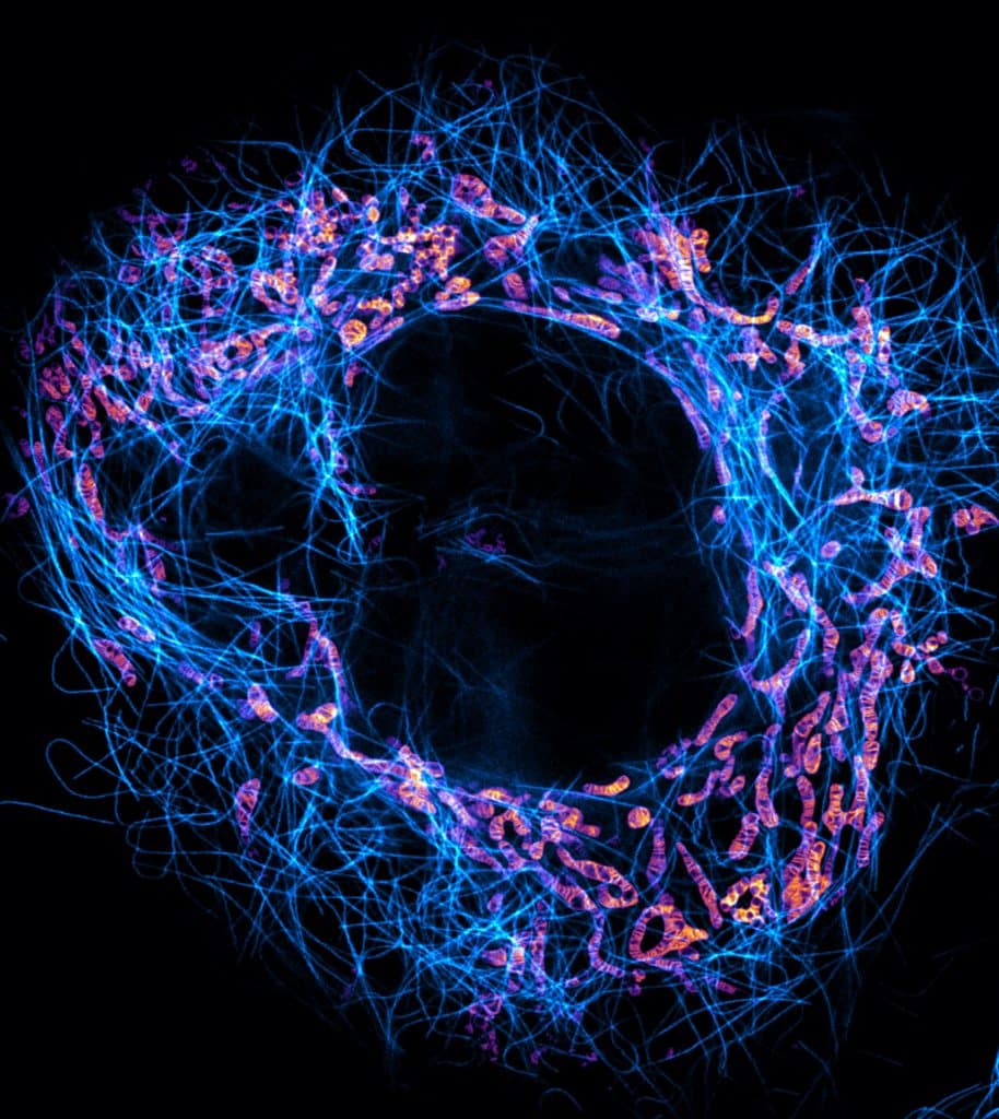
Our featured image, acquired by Till Stephan, shows a live-cell stimulated emission depletion (STED) microscopy recording of a HeLa cell in which mitochondria (orange-magenta) and microtubules (cyan) are labelled. The super-resolution image reveals the densely stacked mitochondrial cristae, coordinated invaginations of the mitochondrial inner membrane.
We caught up with Till to find out about his research and what he is excited about in microscopy.
Research career so far: I completed my biochemistry studies in 2015 at the Hannover Medical School (MHH), where I conducted research on actin cytoskeleton dynamics under the guidance of Professor Jan Faix. Driven by my passion for light microscopy, I pursued my PhD at the Max Planck Institute for Biophysical Chemistry in Göttingen, joining the esteemed department led by Professor Stefan Hell in 2016. During my doctoral studies, I had the privilege of working in the research group of Professor Stefan Jakobs, where I focused on investigating the organization of the mitochondrial inner membrane using super-resolution microscopy techniques. I successfully defended my PhD in 2020 and have since continued my scientific journey as a postdoctoral researcher in the same institute.
Current research: Throughout my PhD, our research group, alongside other research groups, has uncovered remarkable dynamics of the mitochondrial inner membrane, particularly focusing on the intriguing dynamic behaviour of mitochondrial cristae. Presently, our objective is to gain a deeper understanding of the underlying factors that govern these dynamics. To achieve this, we collaborate with chemists and physicists to develop innovative tools that enable us to observe mitochondrial cristae dynamics with exceptional precision and minimal phototoxicity.
Favourite imaging technique/microscope: My favourite imaging technique is STED microscopy due to its ability to provide super-resolved images without the need for image reconstruction. Additionally, this technique offers the advantage of being relatively fast, enabling the observation of dynamic, living systems.
What are you most excited about in microscopy? I am excited about the ever-expanding applications of super-resolution microscopy, fuelled by the continuous development of new advanced labelling strategies. This remarkable progress has paved the way for exploring entirely new biological questions. Furthermore, the relentless efforts of optical engineers and physicists are consistently pushing the frontiers of light microscopy, opening up new possibilities and horizons.
 (No Ratings Yet)
Dr. Stephan, your images are amazing. How might I obtain a copy of the image published in Science Focus Magazine of the actin-based protein structures within cells that form sarcomeres. Thank you for doing this wonderful and impressive work.