Featured image with Nick Gatford
Posted by FocalPlane, on 16 November 2023
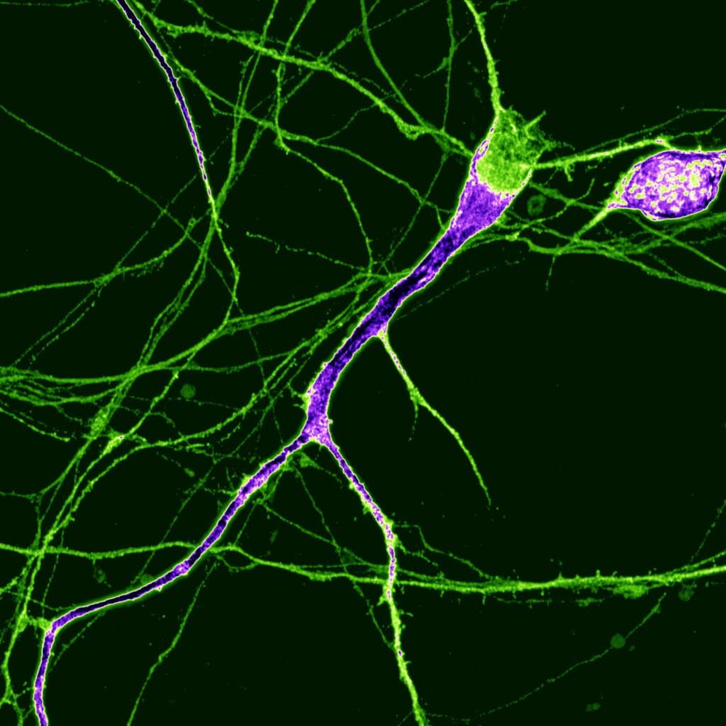
Our featured image shows a single human dopaminergic neuron generated from a human stem cell, acquired via super-resolution Airyscan confocal microscopy at the University of Oxford Micron facility by Nick Gatford.
We caught up with Nick to find out about his research and what he is excited about in microscopy.
Research career so far: I completed my PhD in Neuroscience at King’s College London where I was first inspired to pursue microscopy as a core methodology of my PhD project. I was particularly keen on using super-resolution microscopy to better understand how autism-associated cell adhesion molecules influence early stages of neurodevelopment in human stem cell-derived neurons. This led to my first primary research paper, in which I especially aimed to show the subcellular phenotype in as fine detail and resolution as possible: https://doi.org/10.1093/hmg/ddab277. Some of the most memorable research papers, presentations, and posters I’ve seen have taken Anton Chekov’s ‘show, don’t tell’ approach to heart and shown the audience what they’ve discovered with a microscopy image rather than telling the audience with a wall of text or verbally. I’ve since learnt to adopt this approach wherever possible and I actively encourage my students to do the same.
Current research: Since completing my PhD, I have taken up a postdoctoral research associate position at the University of Oxford Kavli Institute for Nanoscience Discovery, where I am currently applying my microscopy skills to better understand how Parkinson’s disease develops using human stem cell-derived dopaminergic neurons. This is primarily focused on the main pathogenic component of Parkinson’s, alpha-synuclein, and its most critical interacting partners when it pathologically aggregates in dopaminergic neurons. The microscopes in the Micron Imaging Facility here at Oxford are world-class and have already been instrumental to my work, hopefully leading to new discoveries about how this devastating disease emerges and ultimately how we can prevent these neurons from degenerating in human patients.
Favourite imaging technique/microscope: That’s definitely a tricky one! It seems every few months there’s an exciting new microscopy technique to learn about as the field advances towards more bespoke microscope builds and image analysis tools. Much of my PhD was spent using an iSIM system and several of the Micron microscopes are SIM set ups, so SIM is certainly a contender. That being said, I’m always blown away by the resolution of single molecule techniques like dSTORM or PAINT, especially when looking at highly localised individual synaptic proteins at the nanoscopic level. Ultimately though I would have to say I enjoy any method that lets me see the intricate details of mammalian neurobiology, particularly if it means seeing something down a microscope that no one else has seen before.
What are you most excited about in microscopy? Cautiously, I would say AI and how it could potentially be used in all sorts of microscopy applications like detecting patterns in thousands of microscopy images (nuclei, puncta, subcellular structures etc), whole brain tissue connectomics, or even simple denoising. Its potential to revolutionise all areas of biology are huge, especially at the systems level. That being said, giants of the AI field Demis Hassabis and Geoffrey Hinton have both recently urged the AI community for a more measured approach to developing new AI systems thanks to the equally substantial potential to do more harm than good if not used for the right reasons.
For updates on what the Tofaris lab is up to and more microscopy images, please follow their recently opened X (Twitter) account: @tofarislab
 (No Ratings Yet)