Microscopy preprints – New tools and techniques
Posted by FocalPlane, on 8 October 2021
Here is a curated selection of preprints published recently. In this post, we focus specifically on new imaging tools only.
Preprint selected by Pablo J. Sáez who also participated in our Hot Reads post this month:
A turquoise fluorescence lifetime-based biosensor for quantitative imaging of intracellular calcium. Franka H. van der Linden, Eike K. Mahlandt, Janine J.G. Arts, VJoep Beumer, Jens Puschhof, Saskia M.A. de Man, Anna O. Chertkova, Bas Ponsioen, Hans Clevers, Jaap D. van Buul, Marten Postma, Theodorus W.J. Gadella Jr., Joachim Goedhart
By engineering a fluorescent protein (mTurquoise2), the authors develop a new calcium sensor. This sensor, named Tq-Ca-FLITS, detects calcium and reports with a change in the fluorescence lifetime, which can be calibrated and subsequently used to the measure absolute calcium concentrations. Tq-Ca-FLITS can be targeted to different organelles.
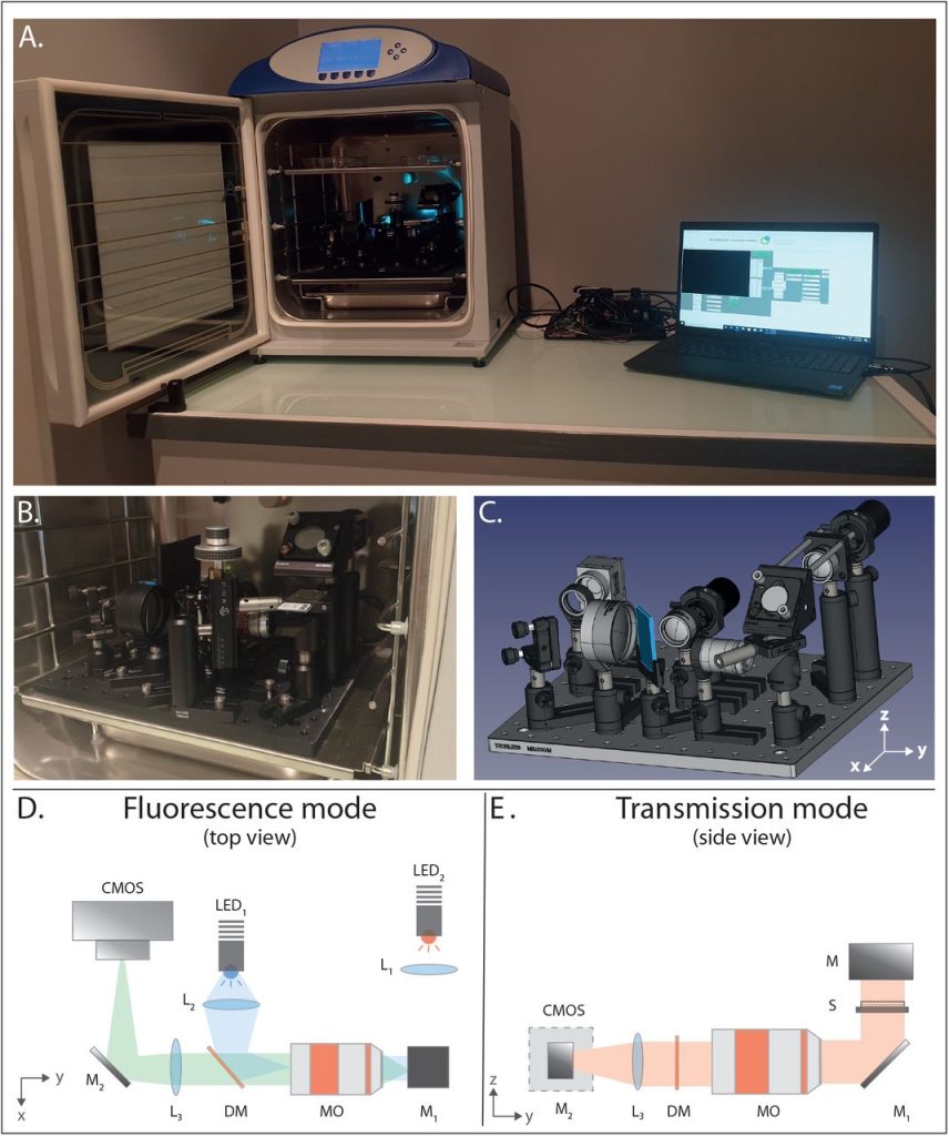
The Incubascope : a simple, compact and large field of view microscope for long-term imaging inside an incubator. Amaury Badon, Laetitia Andrique, Amaël Mombereau, Louis Rivet, Adeline Boyreau, Pierre Nassoy, Gaëlle Recher
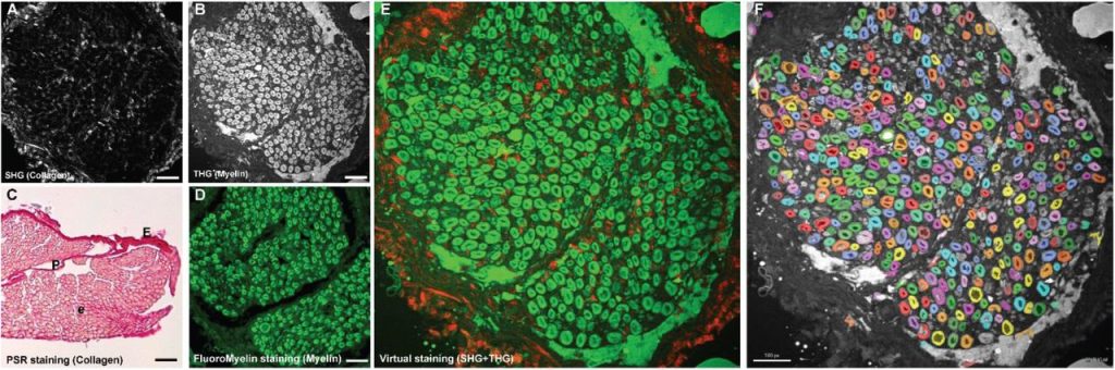
Multiphoton microscopy for label-free multicolor imaging of peripheral nerve. Lars Rishøj, Iván Coto Hernández, Siddharth Ramachandran, Nate Jowett
Determination of protein stoichiometries via dual-color colocalization with single molecule localization microscopy. Hua Leonhard Tan, Stefanie Bungert-Plümke, Daniel Kortzak, Christoph Fahlke, Gabriel Stölting
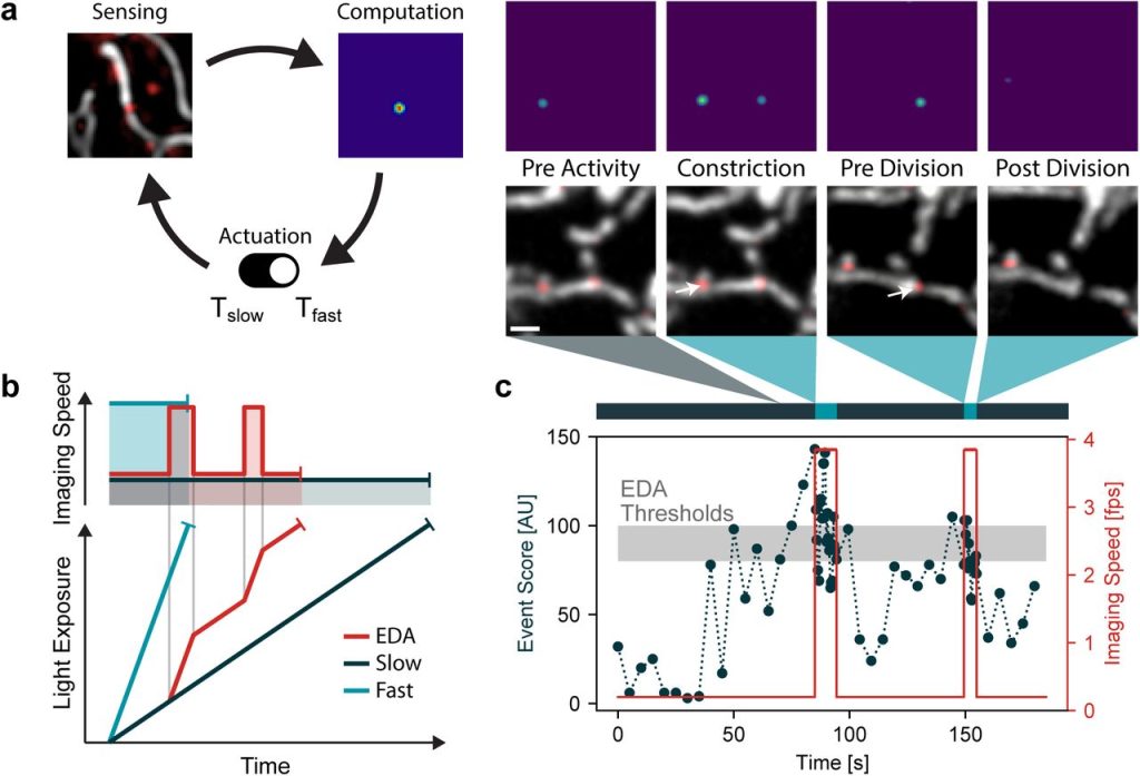
Event-driven acquisition for content-enriched microscopy. Dora Mahecic, Willi L. Stepp, Chen Zhang, Juliette Griffié, Martin Weigert, Suliana Manley
Acoustic light-sheet microscopy. S. Wunderl, A. Ishijima, E. A. Susaki, Z. Xu, H. Song, H. Zha, T. Azuma, I. Sakuma, H. Fukuoka, E. Okada, H. R. Ueda, S. Takagi, K. Nakagawa
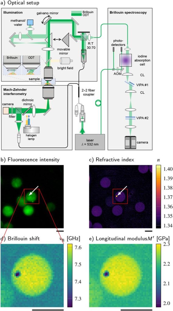
Combined fluorescence, optical diffraction tomography and Brillouin microscopy.
Raimund Schlüßler, Kyoohyun Kim, Martin Nötzel, Anna Taubenberger, Shada Abuhattum, Timon Beck, Paul Müller, Shovamayee Maharana, Gheorghe Cojoc, Salvatore Girardo, Andreas Hermann, Simon Alberti, Jochen Guck
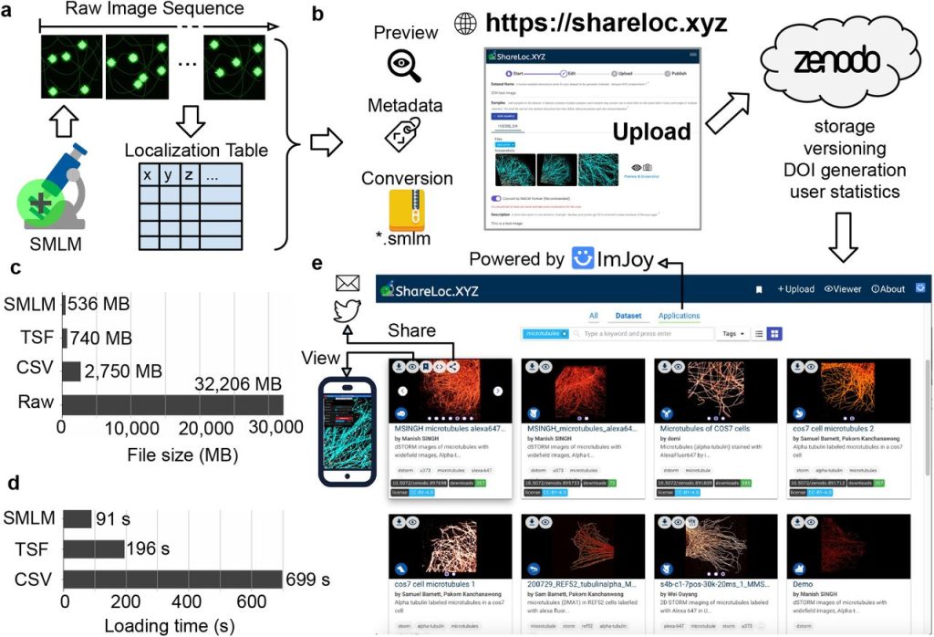
ShareLoc – an open platform for sharing localization microscopy data. Jiachuan Bai, Wei Ouyang, Manish Kumar Singh, Christophe Leterrier, Paul Barthelemy, Samuel F.H. Barnett, Teresa Klein, Markus Sauer, Pakorn Kanchanawong, Nicolas Bourg, Mickael M. Cohen, Benoît Lelandais, Christophe Zimmer
Versatile, do-it-yourself, low-cost spinning disk confocal microscope. Aaron R. Halpern, Min Yen Lee, Marco D. Howard, Marcus A. Woodworth, Philip R. Nicovich, Joshua C. Vaughan
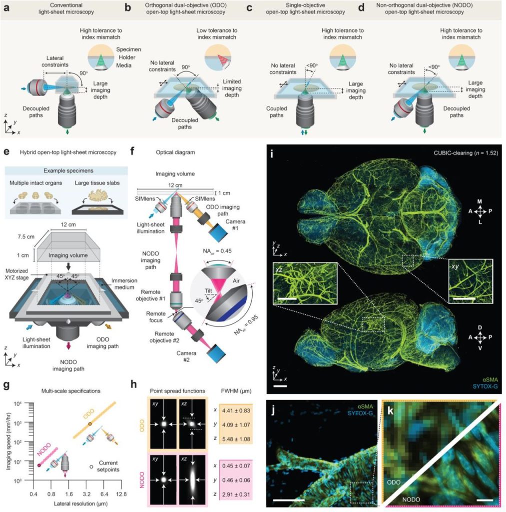
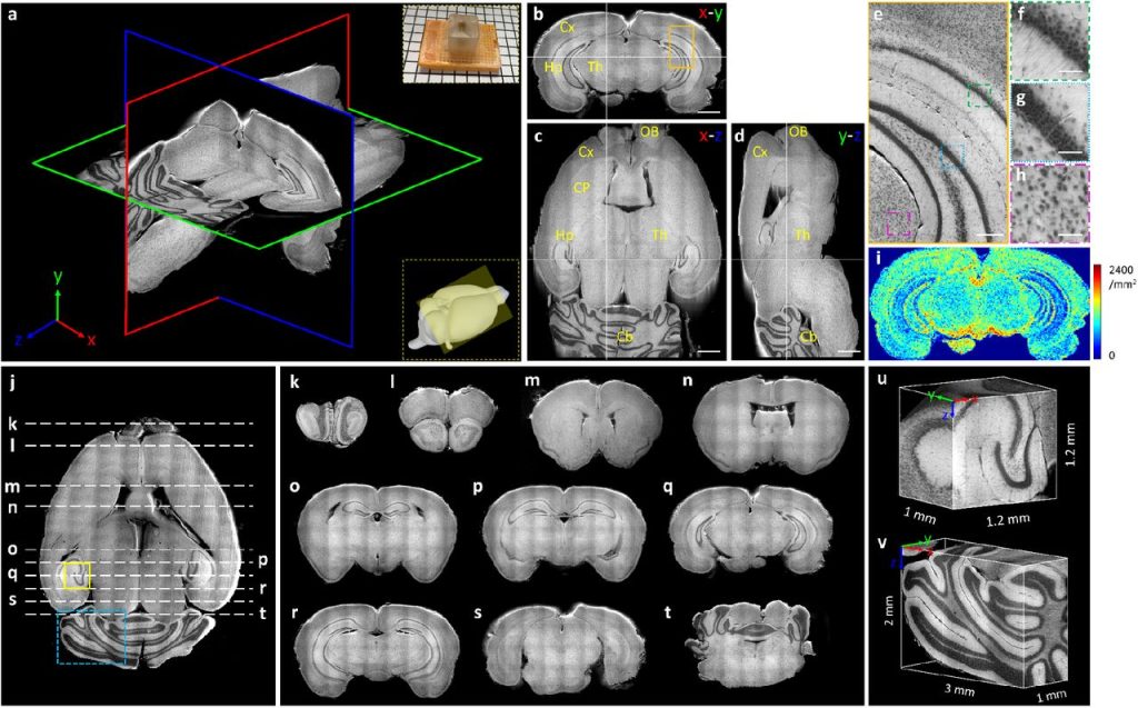
A hybrid open-top light-sheet microscope for multi-scale imaging of cleared tissues. Adam K. Glaser, Kevin W. Bishop, Lindsey A. Barner, Etsuo A. Susaki, Shimpei I. Kubota, Gan Gao, Robert B. Serafin, Pooja Balaram, Emily Turschak, Philip R. Nicovich, Hoyin Lai, Luciano A.G. Lucas, Yating Yi, Eva K. Nichols, Hongyi Huang, Nicholas P. Reder, Jasmine J. Wilson, Ramya Sivakumar, Elya Shamskhou, Caleb R. Stoltzfus, Xing Wei, Andrew K. Hempton, Marko Pende, Prayag Murawala, Hans U. Dodt, Takato Imaizumi, Jay Shendure, Brian J. Beliveau, Michael Y. Gerner, Li Xin, Hu Zhao, Lawrence D. True, R. Clay Reid, Jayaram Chandrashekar, Hiroki R. Ueda, Karel Svoboda, Jonathan T.C. Liu
Two-color super-resolution localization microscopy via joint encoding of emitter location and color. Yujie Wang, Weibing Kuang, Mingtao Shang, Zhen-Li Huang
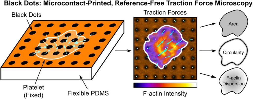
Black Dots: Microcontact-Printed, Reference-Free Traction Force Microscopy. Kevin M. Beussman, Molly Y. Mollica, Andrea Leonard, Jeffrey Miles, John Hocter, Zizhen Song, Moritz Stolla, Sangyoon J. Han, Ashley Emery, Wendy E. Thomas, Nathan J. Sniadecki
Systematic Transmission Electron Microscopy-Based Identification of Cellular Degradation Machinery. Kit Neikirk, Zer Vue, Prasanna Katti, Jianqiang Shao, Trace Christensen, Edgar Garza Lopez, Andrea Marshall, Caroline B. Palavicino-Maggio, Jessica Ponce, Ahmad Alghanem, Larry Vang, Heather K. Beasley, Taylor Rodman, Margaret Mungai, Marcelo Correia, Vernat Exil, Sandra A. Murray, Jeffrey L. Salisbury, Brian Glancy, Renata O. Pereira, E. Dale Abel, Antentor O. Hinton Jr.
Single-Molecule Localization Microscopy of 3D Orientation and Anisotropic Wobble using a Polarized Vortex Point Spread Function. Tianben Ding, Matthew D. Lew
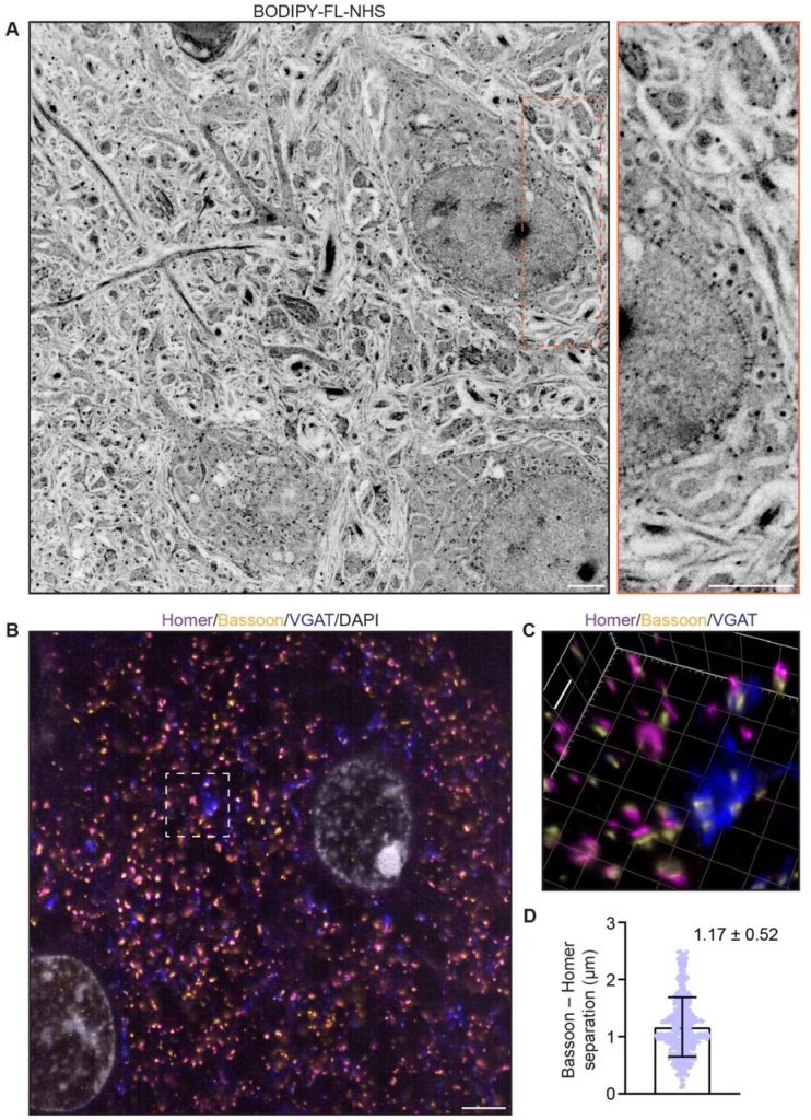
Visualizing cellular and tissue ultrastructure using Ten-fold Robust Expansion Microscopy (TREx)
Hugo G.J. Damstra, Boaz Mohar, Mark Eddison, Anna Akhmanova, Lukas C. Kapitein, Paul W. Tillberg
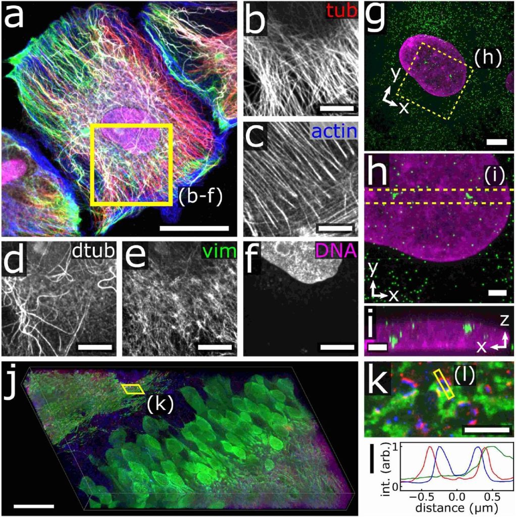
How to Extend the Capabilities of a Commercial Two-Photon Microscope to Perform Super-Resolution Imaging, Wavelength Mixing and Label-Free Microscopy. Chiara Peres, Chiara Nardin, Guang Yang, Fabio Mammano
Three-dimensional label-free histological imaging of whole organs by microtomy-assisted autofluorescence tomography. Yan Zhang, Lei Kang, Wentao Yu, Victor Tsz Chun Tsang, Terence T. W. Wong
A technique for resolution assessment in blind-SIM experiments. Imen Boujmil, Emmanouil Xypakis, Giancarlo Ruocco, Marco Leonetti
 (No Ratings Yet)
One thought on “Microscopy preprints – New tools and techniques”
Leave a Reply
Get involved
Create an account or log in to post your story on FocalPlane.
More posts like this

Filter by
- NewsApply
- DiscussionsApply
- How toApply
- ToolsApply
- Case studiesApply
- InterviewsApply
- JobsApply
- EducationApply
- Blog seriesApply
- Volume EMApply
- Latin American Micro..scopistsApply
- Bio-image Analysis w..ith NapariApply
- Imaging with…Apply
- Towards Global Acces..sApply
- Latin America Bioima..gingApply
- From Zero to Qupath ..HeroApply
- Asian Microscopists ..and Cell BiologistsApply
- AIC at HHMI JaneliaApply
- Deep Learning for Bi..o-image analysisApply
- GloBIAS – updates fr..om the communityApply
- WAMBIAN: West Africa.. in FocusApply
- Highlights from Euro..-BioImagingApply
- LSFM seriesApply
- DIY MicroscopyApply
- View all
Very interesting. Amazing photos