Microscopy preprints – applications in cell biology and more
Posted by FocalPlane, on 22 October 2021
Here is a curated selection of preprints published recently. In this post, we focus specifically on preprints using microscopy tools in different fields such as cell biology, neuroscience, and development.
These two preprints were selected by Pablo J. Sáez who also participated in our Hot Reads post this month:
An anti-inflammatory activation sequence governs macrophage transcriptional dynamics during tissue injury. Nicolas Denans, Nhung T. T. Tran, Madeleine E. Swall, Daniel C. Diaz, Jillian Blanck, Tatjana Piotrowski
“Using beautiful in vivo imaging in zebrasifsh, the authors show that macrophage are sequentially activated to migrate towards the injury site and resolve inflammation. Elegant use of photoconversion to show that it is the same population of macrophages that perform this function.”
Mechanical intercellular communication via matrix-borne cell force transmission during vascular network formation. Christopher D. Davidson, Samuel J. DePalma, William Y. Wang, Jordan L. Kamen, Danica Kristen P. Jayco, Brendon M. Baker
“The authors show that mechanical intercellular communication between endothelial cells depends on calcium signaling and focal adhesion by using hydrogels. Cellular alignment of the fibers allows force transmission and cluster formation.”
A cellular and molecular atlas reveals the basis of chytrid development. Davis Laundon, Nathan Chrismas, Kimberley Bird, Seth Thomas, Thomas Mock, Michael Cunliffe
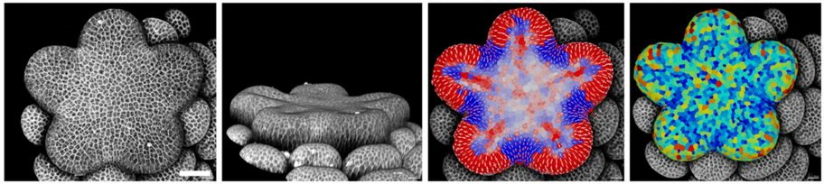
Quantitative live-imaging of Aquilegia floral meristems reveals distinct patterns of floral organ initiation and cell-level dynamics of floral meristem termination. Ya Min, Stephanie J. Conway, Elena M. Kramer
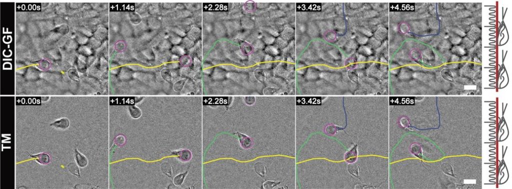
High definition DIC imaging uncovers transient stages of pathogen infection cycles on the surface of human adult stem cell-derived intestinal epithelium. Jorik M. van Rijn, Jens Eriksson, Jana Grüttner, Magnus Sundbom, Dominic-Luc Webb, Per M. Hellström, Staffan G. Svärd, Mikael E. Sellin
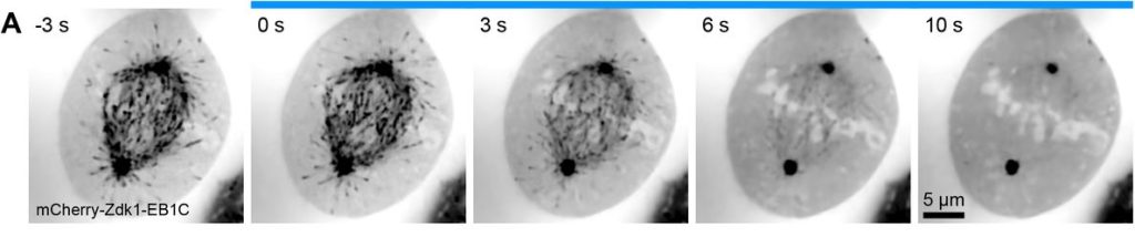
Optogenetic EB1 inactivation shortens metaphase spindles by disrupting cortical force-producing interactions with astral microtubules. Alessandro Dema, Jeffrey van Haren, Torsten Wittmann
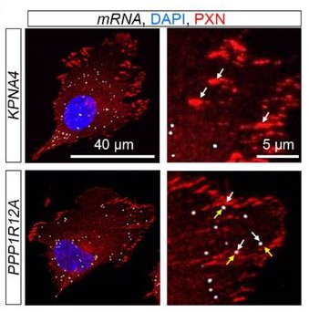
Non-coding function for mRNAs in Focal Adhesion Architecture and Mechanotransduction. Liana Boraas, Mengwei Hu, Lauren Thornton, Charles E. Vejnar, Gang Zhen, Antonio J. Giraldez, Christine Mayr, Siyuan Wang, Stefania Nicoli
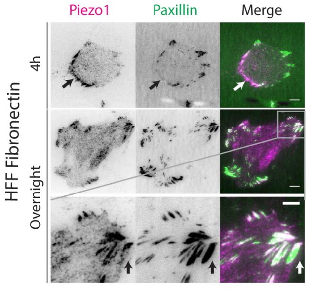
Force-dependent Piezo1 recruitment to focal adhesions regulates adhesion maturation and turnover specifically in non-transformed cells. Mingxi Yao, Ajay Tijore, Charles D Cox, Anushya Hariharan, Guy Tran Van Nhieu, Boris Martinac, Michael Sheetz
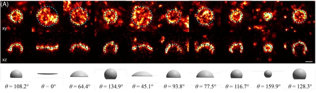
Superresolution microscopy reveals partial preassembly and subsequent bending of the clathrin coat during endocytosis. Markus Mund, Aline Tschanz, Yu-Le Wu, Felix Frey, Johanna L. Mehl, Marko Kaksonen, Ori Avinoam, Ulrich S. Schwarz, Jonas Ries
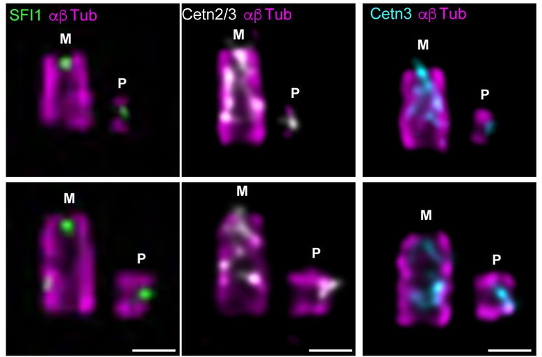
SFI1 and centrin form a distal end complex critical for proper centriole architecture and ciliogenesis. Imène B. Bouhlel, Marine. H. Laporte, Eloïse Bertiaux, Alexia Giroud, Susanne Borgers, Juliette Azimzadeh, Michel Bornens, Paul Guichard, Anne Paoletti, Virginie Hamel
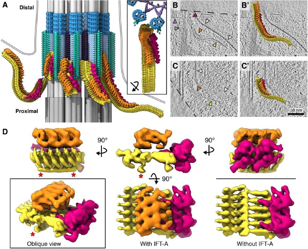
In situ architecture of the ciliary base reveals the stepwise assembly of IFT trains. Hugo van den Hoek, Nikolai Klena, Mareike A. Jordan, Gonzalo Alvarez Viar, Miroslava Schaffer, Philipp S. Erdmann, William Wan, Jürgen M. Plitzko, Wolfgang Baumeister, Gaia Pigino, Virginie Hamel, Paul Guichard, Benjamin D. Engel
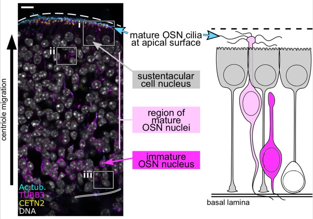
Long-range migration of centrioles to the apical surface of the olfactory epithelium. Kaitlin Ching, Jennifer T. Wang, Tim Stearns
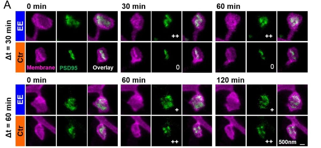
Environmental enrichment enhances patterning and remodeling of synaptic nanoarchitecture revealed by STED nanoscopy. Waja Wegner, Heinz Steffens, Carola Gregor, Fred Wolf, Katrin I. Willig.
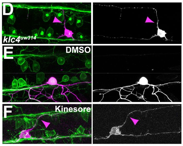
KLC4 shapes axon arbors during development and mediates adult behavior. Elizabeth M. Haynes, Jiaye “Henry” He, Marcel Jean-Pierre, Kevin W. Eliceiri, Jan Huisken, Mary C. Halloran
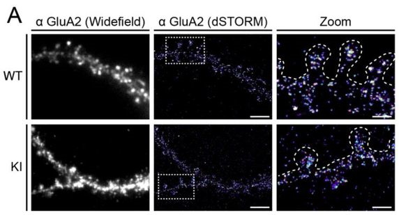
High-resolution imaging and manipulation of endogenous AMPA receptor surface mobility during synaptic plasticity and learning. Angela M. Getz, Mathieu Ducros, Christelle Breillat, Aurélie Lampin-Saint-Amaux, Sophie Daburon, Urielle François, Agata Nowacka, Mónica Fernández-Monreal, Eric Hosy, Frédéric Lanore, Hanna Zieger, Matthieu Sainlos, Yann Humeau, Daniel Choquet
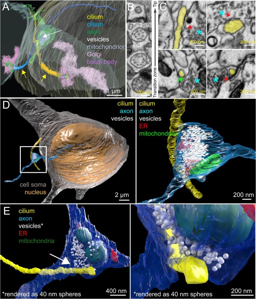
A serotonergic axon-cilium synapse drives nuclear signaling to maintain chromatin accessibility.
Shu-Hsien Sheu, Srigokul Upadhyayula, Vincent Dupuy, Song Pang, Andrew L. Lemire, Deepika Walpita, H. Amalia Pasolli, Fei Deng, Jinxia Wan, Lihua Wang, Justin Houser, Silvia Sanchez-Martinez, Sebastian E. Brauchi, Sambashiva Banala, Melanie Freeman, C. Shan Xu, Tom Kirchhausen, Harald F. Hess, Luke Lavis, Yu-Long Li, Séverine Chaumont-Dubel, David E. Clapham
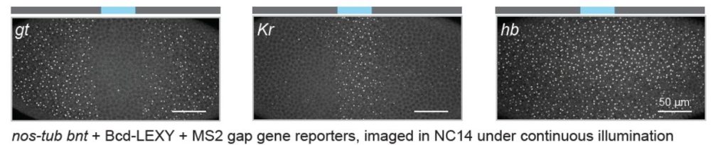
Optogenetic control of the Bicoid morphogen reveals fast and slow modes of gap gene regulation.
Anand P. Singh, Ping Wu, Sergey Ryabichko, João Raimundo, Michael Swan, Eric Wieschaus, Thomas Gregor, Jared E. Toettcher
 (No Ratings Yet)
Get involved
Create an account or log in to post your story on FocalPlane.
More posts like this

Filter by
- NewsApply
- DiscussionsApply
- How toApply
- ToolsApply
- Case studiesApply
- InterviewsApply
- JobsApply
- EducationApply
- Blog seriesApply
- WAMBIAN: West Africa.. in FocusApply
- Volume EMApply
- Latin American Micro..scopistsApply
- Bio-image Analysis w..ith NapariApply
- Imaging with…Apply
- Towards Global Acces..sApply
- Latin America Bioima..gingApply
- From Zero to Qupath ..HeroApply
- Asian Microscopists ..and Cell BiologistsApply
- AIC at HHMI JaneliaApply
- Deep Learning for Bi..o-image analysisApply
- GloBIAS – updates fr..om the communityApply
- Highlights from Euro..-BioImagingApply
- LSFM seriesApply
- DIY MicroscopyApply
- View all