Microscopy preprints – Bioimage analysis tools
Posted by FocalPlane, on 22 April 2022
Here is a curated selection of preprints published recently. In this post, we focus specifically on new bioimage analysis tools only.
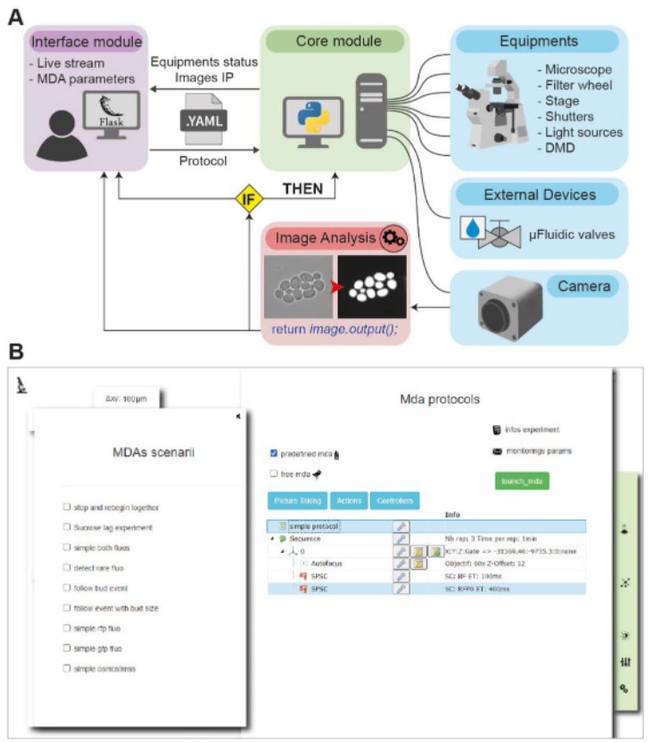
CyberSco.py: open-source software for event-based, conditional microscopy
Lionel Chiron, Matthias LeBec, Celine Cordier, Sylvain Pouzet, Dimitrije Milunov, AlvaroBanderas, Jean-Marc di Meglio, Benoit Sorre, Pascal Hersen
LiveCellMiner: A New Tool to Analyze Mitotic Progression
Daniel Moreno-Andres, Anuk Bhattacharyya, Anja Scheufen, Johannes Stegmaier
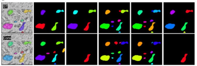
Ryan Conrad, Kedar Narayan
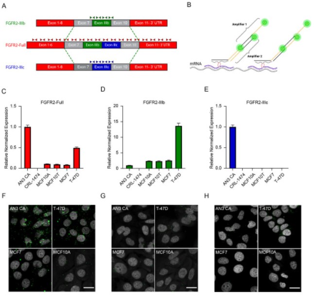
HiFENS: High-throughput FISH detection of endogenous pre-mRNA splicing isoforms
Asaf Shilo, Gianluca Pegoraro, Tom Misteli
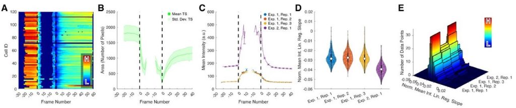
Theo Aspert, Didier Hentsch, Gilles Charvin
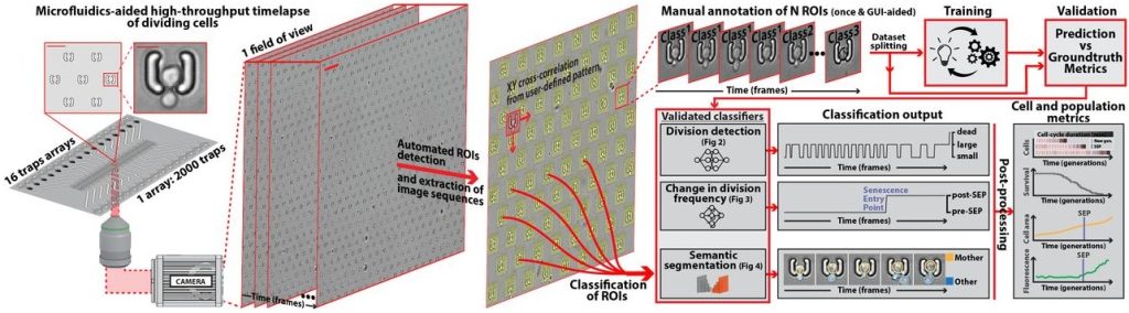
Figure extracted from Aspert et al.
Celine Trebeau, Jacques Boutet de Monvel, Gizem Altay, Jean-Yves Tinevez, Raphael Etournay
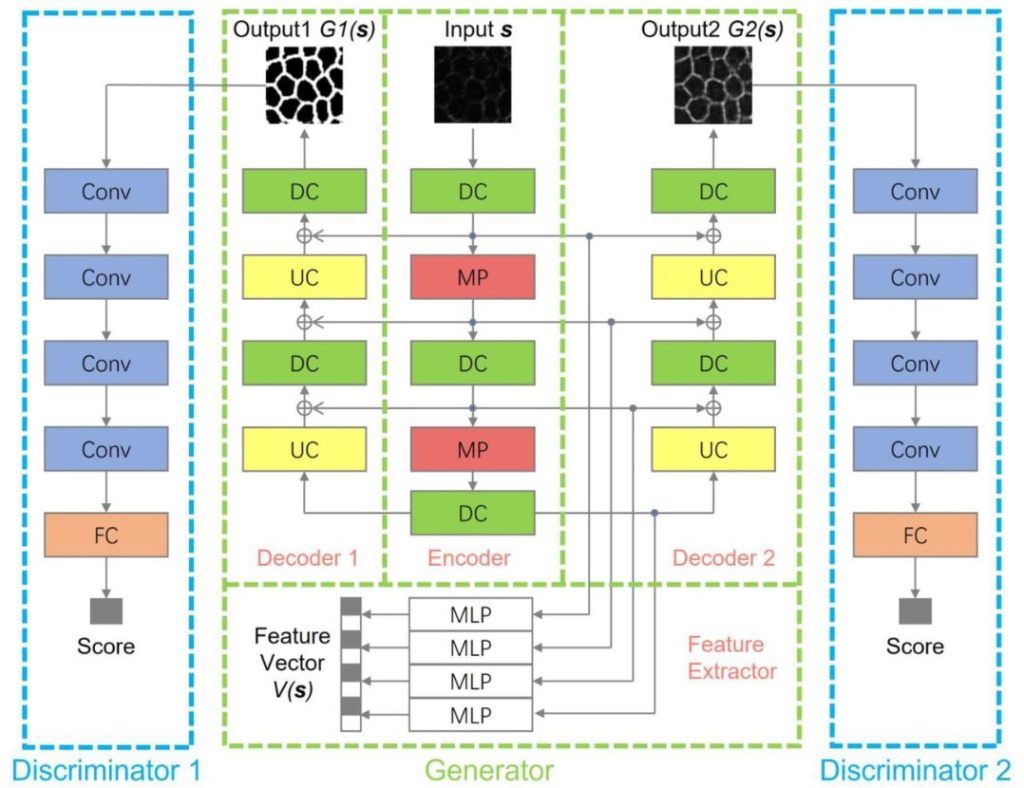
Ankit Gupta, Alan Sabirsh, Carolina Wahlby, Ida-Maria Sintorn
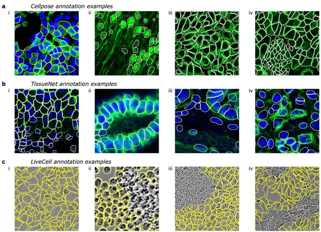
Cellpose 2.0: how to train your own model
Carsen Stringer, Marius Pachitariu
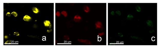
Abbas Habibalahi, Jared Campbell, Saabah Mahbub, Ayad Anwer, Long Nguyen, Anthony Gill, Muh Wong, Angela Chou, Carol Pollock, Sonia Saad, Ewa Goldys
Hanyi Yu, Fusheng Wang, George Theodoro, John Nickerson, Jun Kong
 (1 votes, average: 1.00 out of 1)