Microscopy preprints – new tool and techniques in imaging and bioimage analysis
Posted by FocalPlane, on 5 December 2022
Here is a curated selection of preprints published recently. This post is a bumper edition, featuring preprints on bioimage analysis and new tools and techniques in imaging.
Bioimage analysis
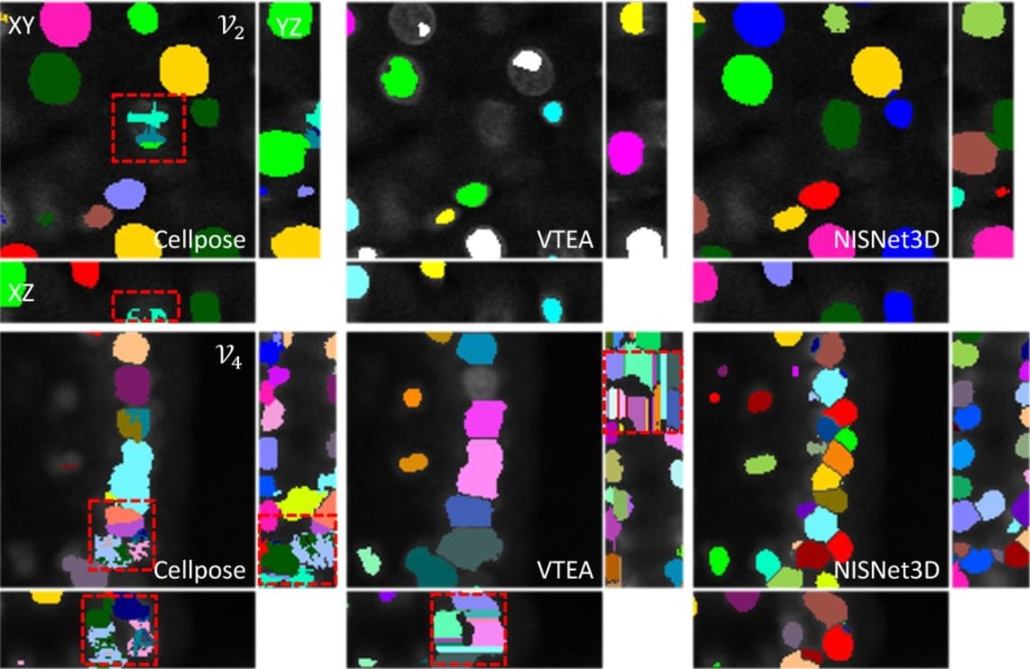
NISNet3D: Three-Dimensional Nuclear Synthesis and Instance Segmentation for Fluorescence Microscopy Images
ProfileLiming Wu, ProfileAlain Chen, ProfilePaul Salama, ProfileKenneth Dunn, ProfileEdward Delp
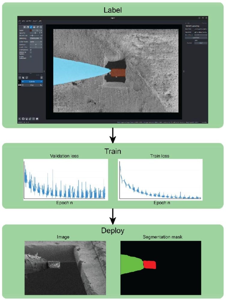
Deep neural network automated segmentation of cellular structures in volume electron microscopy
ProfileBenjamin Gallusser, ProfileGiorgio Maltese, ProfileGiuseppe Di Caprio, ProfileTegy John Vadakkan, ProfileAnwesha Sanyal, Elliott Somerville, ProfileMihir Sahasrabudhe, Justin O’Connor, ProfileMartin Weigert, ProfileTom Kirchhausen
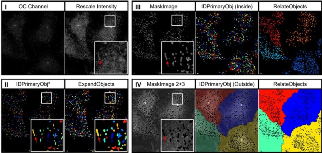
Automated segmentation and quantitative analysis of organelle morphology, localization and content using CellProfiler
ProfileSebastiaan N.J. Laan, Richard J. Dirven, Jeroen Eikenboom, ProfileRuben Bierings, for the SYMPHONY consortium
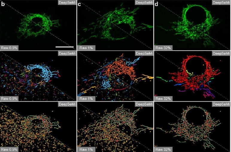
Bio-friendly long-term subcellular dynamic recording by self-supervised image enhancement microscopy
Guoxun Zhang, Xiaopeng Li, Yuanlong Zhang, Xiaofei Han, Xinyang Li, Jinqiang Yu, Boqi Liu, Jiamin Wu, Li Yu, Qionghai Dai
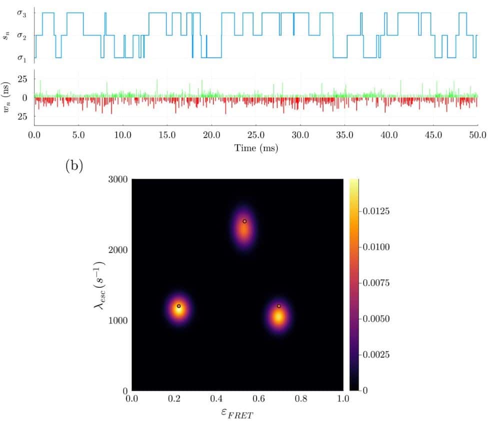
Single Photon smFRET. I. Theory and Conceptual Basis
ProfileAyush Saurabh, ProfileMohamadreza Fazel, Matthew Safar, ProfileIoannis Sgouralis, ProfileSteve Pressé
Single Photon smFRET. II. Application to Continuous Illumination
ProfileAyush Saurabh, Matthew Safar, ProfileMohamadreza Fazel, ProfileIoannis Sgouralis, Steve Pressé
Single Photon smFRET. III. Application to Pulsed Illumination
Matthew Safar, ProfileAyush Saurabh, ProfileBidyut Sarkar, ProfileMohamadreza Fazel, ProfileKunihiko Ishii, ProfileTahei Tahara, ProfileIoannis Sgouralis, ProfileSteve Pressé
Label-free cleared tissue microscopy and machine learning for 3D histopathology of biomaterial implants
Tran B Ngo, Sabrina DeStefano, Jiamin Liu, Yijun Su, Hari Shroff, Harshad D Vishwasrao, Kaitlyn Sadtler
CellSighter – A neural network to classify cells in highly multiplexed images
Yael Amitay, ProfileYuval Bussi, Ben Feinstein, ProfileShai Bagon, ProfileIdan Milo, ProfileLeeat Keren
Root Walker: an automated pipeline for large scale quantification of early root growth responses at high spatial and temporal resolution
Platre Matthieu Pierre, Halvorson Zachary, Mehta Preyanka, Brent Lukas, Gleason F. Matias, Faizi Kian, ProfileBusch Wolfgang
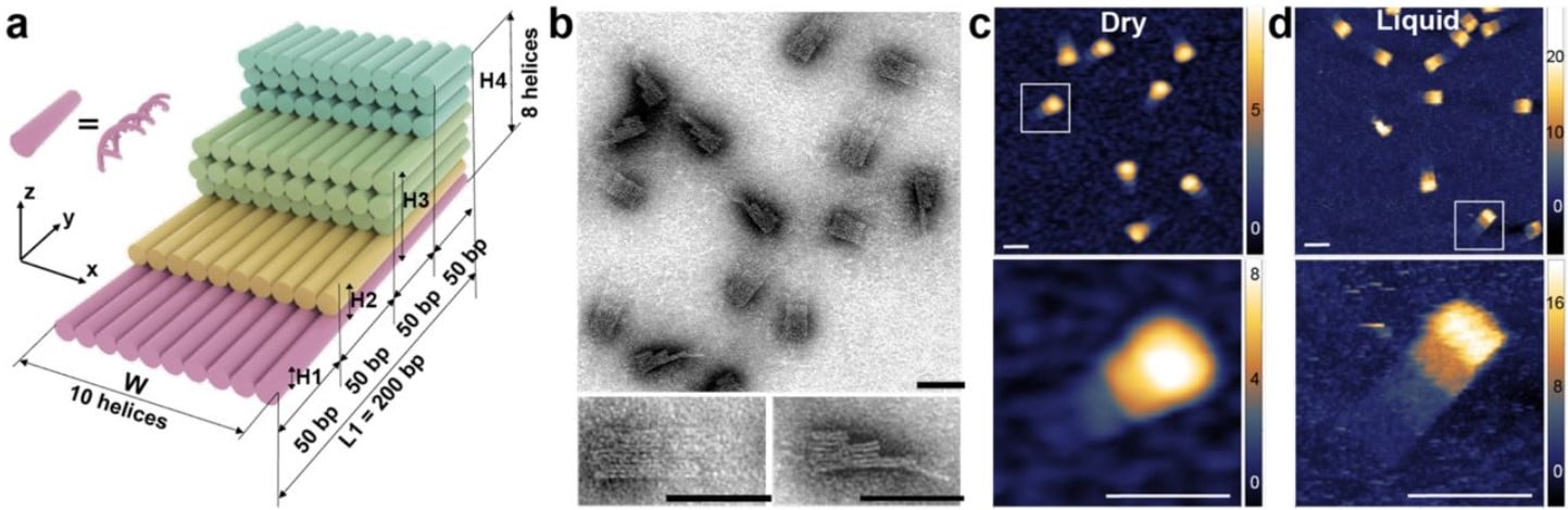
A DNA origami fiducial for accurate 3D AFM imaging
Pauline J. Kolbeck, Mihir Dass, Irina V. Martynenko, Relinde J.A. van Dijk-Moes, Kelly J.H. Brouwer, Alfons van Blaaderen, Willem Vanderlinden, Tim Liedl, ProfileJan Lipfert
Simultaneous Noise Reduction and Layer Segmentation for Visible Light Optical Coherence Tomography in Human Retina
Tianyi Ye, Jingyu Wang, Ji Yi

Multi-site assessment of reproducibility in high-content live cell imaging data
Jianjiang Hu, Xavier Serra-Picamal, Gert-Jan Bakker, Marleen Van Troys, Sabina Winograd-katz, Nil Ege, Xiaowei Gong, Yuliia Didan, Inna Grosheva, Omer Polansky, Karima Bakkali, Evelien Van Hamme, Merijn Van Erp, Manon Vullings, Felix Weiss, Jarama Clucas, Anna M. Dowbaj, ProfileErik Sahai, Christophe Ampe, Benjamin Geiger, ProfilePeter Friedl, Matteo Bottai, ProfileStaffan Strömblad
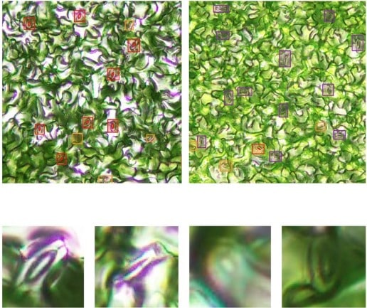
Image-based quantification of Arabidopsis thaliana stomatal aperture from leaf images
Momoko Takagi, Rikako Hirata, Yusuke Aihara, Yuki Hayashi, Miya Mizutani-Aihara, Eigo Ando, Megumi Yoshimura-Kono, Masakazu Tomiyama, Toshinori Kinoshita, Akira Mine, Yosuke Toda
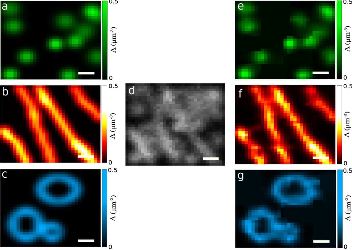
Building Fluorescence Lifetime Maps Photon-by-photon by Leveraging Spatial Correlations
Mohamadreza Fazel, Sina Jazani, Lorenzo Scipioni, Alexander Vallmitjana, Songning Zhu, Enrico Gratton, Michelle A. Digman, Steve Pressé
New tools and techniques
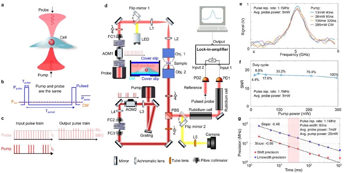
Pulsed stimulated Brillouin microscopy enables high-sensitivity mechanical imaging of live and fragile biological specimens
Fan Yang, Carlo Bevilacqua, Sebastian Hambura, Ana Neves, Anusha Gopalan, Koki Watanabe, Matt Govendir, Maria Bernabeu, Jan Ellenberg, Alba Diz-Muñoz, ProfileSimone Köhler, Georgia Rapti, Martin Jechlinger, ProfileRobert Prevedel
OpenFIBSEM: an application programming interface for easy FIB/SEM automation
Patrick Cleeve, David Dierickx, Genevieve Buckley, Sergey Gorelick, Lucile Naegele, Lachlan Burne, James C Whisstock, ProfileAlex de Marco
Single Pixel Reconstruction Imaging: taking confocal imaging to the extreme
ProfileSimona Streckaitė, Dmitrij Frolov, ProfileJevgenij Chmeliov, ProfileAndrius Gelzinis, Cristian Ilioaia, Sylvie Rimsky, Rienk van Grondelle, ProfileLeonas Valkunas, Andrew Gall, Bruno Robert
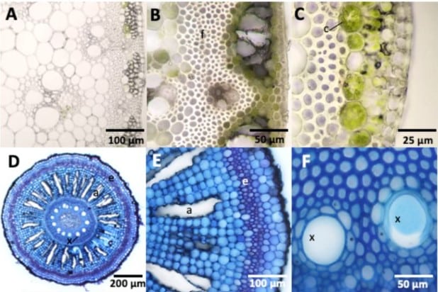
The Rapid-Tome, a 3D-Printed Microtome, and an Updated Hand-Sectioning Method for High-Quality Plant Sectioning
ProfileDavid J. Thomas, Jordan Rainbow, ProfileLaura E. Bartley
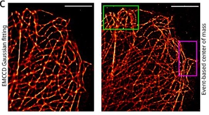
Event-based vision sensor enables fast and dense single-molecule localization microscopy
ProfileClément Cabriel, ProfileChristian G. Specht, ProfileIgnacio Izeddin
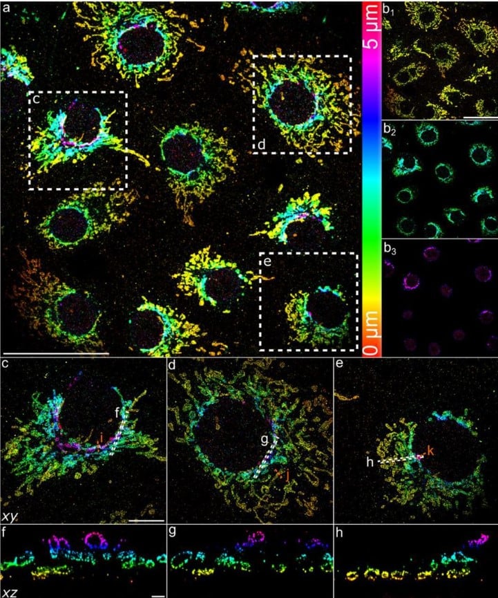
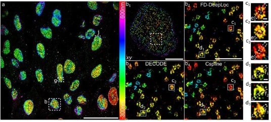
Field dependent deep learning enables high-throughput whole-cell 3D super-resolution imaging
Shuang Fu, Wei Shi, Tingdan Luo, Yingchuan He, Lulu Zhou, Jie Yang, Zhichao Yang, Jiadong Liu, Xiaotian Liu, Zhiyong Guo, Chengyu Yang, Chao Liu, Zhen-li Huang, Jonas Ries, Mingjie Zhang, Peng Xi, Dayong Jin, Yiming Li
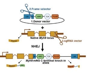
Efficient and rapid fluorescent protein knock-in with universal donors in mammalian stem cells
ProfileYu Shi, Nitya Kopparapu, Lauren Ohler, ProfileDaniel J. Dickinson
Fast-exchanging spirocyclic rhodamine probes for aptamer-based super-resolution RNA imaging
Daniel Englert, Eva-Maria Burger, Jens Lackner, Marko Lampe, Bastian Bühler, Franziska Grün, Janin Schokolowski, ProfileG. Ulrich Nienhaus, ProfileAndres Jäschke, ProfileMurat Sunbul
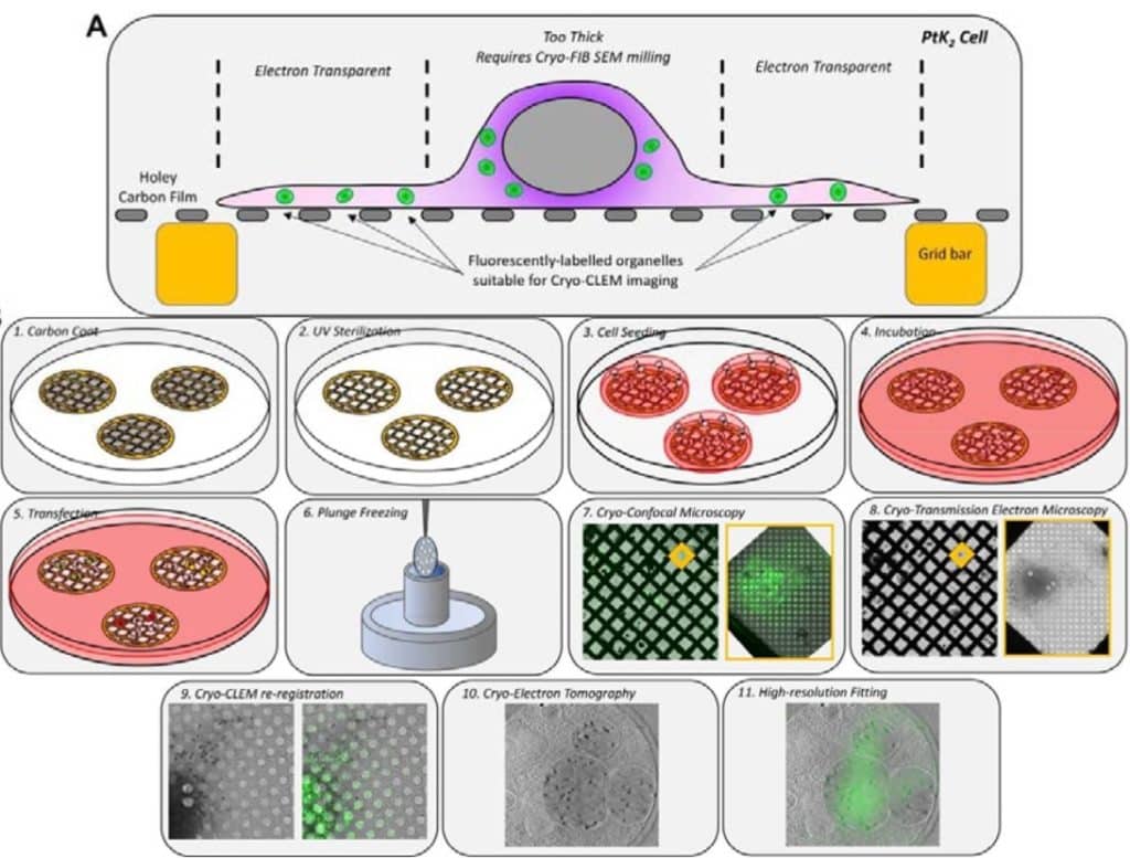
Precision super-resolution cryo-correlative light and electron microscopy for rapid in situ structural analyses of optogenetically-positioned organelles
G.M.I. Redpath, J. Rae, Y. Yao, J. Ruan, M.L. Cagigas, R. Whan, E.C. Hardeman, P.W. Gunning, V. Ananthanarayanan, ProfileR.G. Parton, ProfileN.A. Ariotti
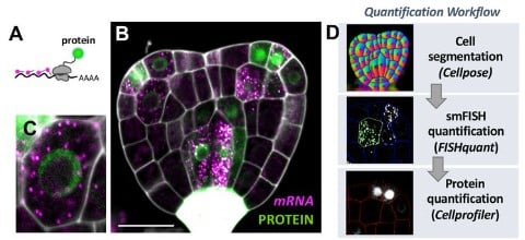
Whole-mount smFISH allows combining RNA and protein quantification at cellular and subcellular resolution
Lihua Zhao, ProfileAlejandro Fonseca, Anis Meschichi, ProfileAdrien Sicard, ProfileStefanie Rosa
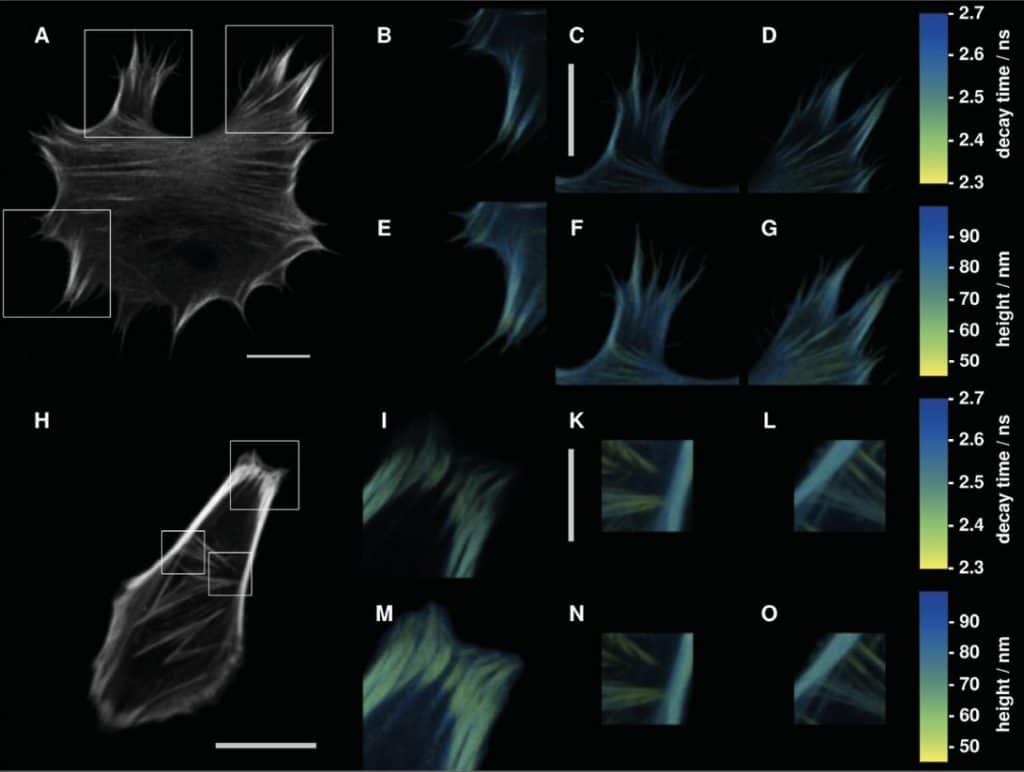
Metal-Induced Energy Transfer (MIET) for Live-Cell Imaging with Fluorescent Proteins
ProfileLara Hauke, ProfileSebastian Isbaner, ProfileArindam Ghosh, ProfileIsabella Guido, Laura Turco, ProfileAlexey I. Chizhik, ProfileIngo Gregor, ProfileNarain Karedla, ProfileFlorian Rehfeldt, ProfileJörg Enderlein
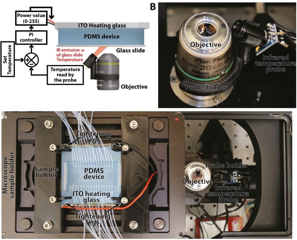
HeatChips: A versatile, low-cost and microscopy-compatible heating system for microfluidic devices
ProfileThéo Aspert, ProfileGilles Charvin
Multiplexed neuropeptide mapping in ant brains integrating microtomography and 3D mass spectrometry imaging
Benedikt Geier, Esther Gil-Mansilla, Zita Liutkeviciute, Roland Hellinger, Jozef Vanden Broeck, Janina Oetjen, ProfileManuel Liebeke, ProfileChristian W. Gruber
Nanoscopy of organelles and tissues with iterative ultrastructure expansion microscopy (iU-ExM)
ProfileVincent Louvel, ProfileRomuald Haase, ProfileOlivier Mercey, ProfileMarine. H. Laporte, ProfileDominique Soldati-Favre, ProfileVirginie Hamel, ProfilePaul Guichard
Field dependent deep learning enables high-throughput whole-cell 3D super-resolution imaging
Shuang Fu, Wei Shi, Tingdan Luo, Yingchuan He, Lulu Zhou, Jie Yang, Zhichao Yang, Jiadong Liu, Xiaotian Liu, Zhiyong Guo, Chengyu Yang, Chao Liu, Zhen-li Huang, Jonas Ries, Mingjie Zhang, Peng Xi, Dayong Jin, Yiming Li
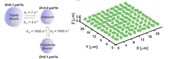
Single Molecule Imaging Simulations with Advanced Fluorophore Photophysics
Dominique Bourgeois
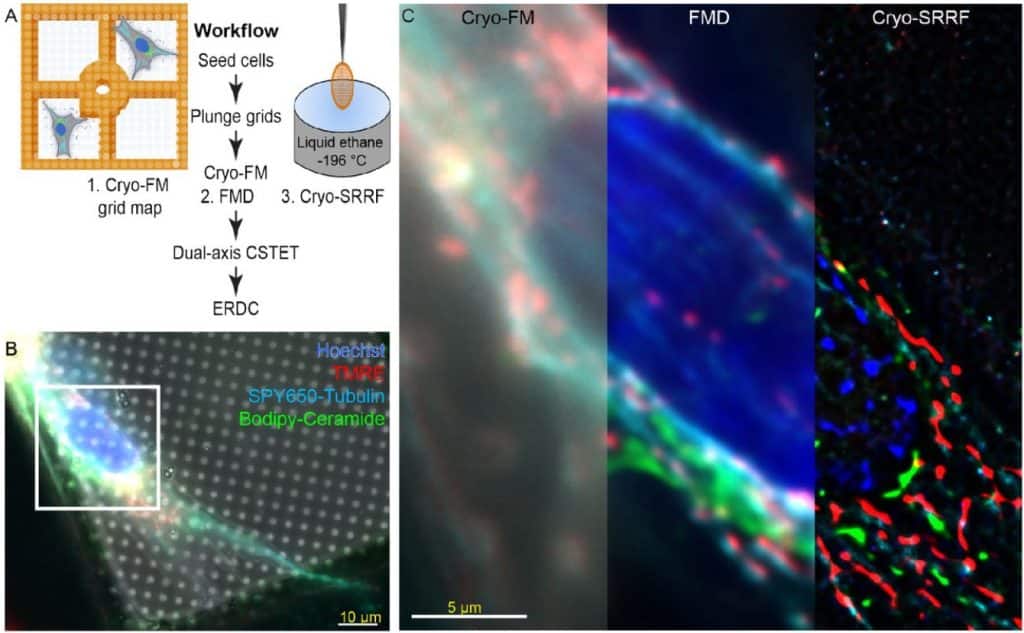
Bridging the light-electron resolution gap with correlative cryo-SRRF and dual-axis cryo-STEM tomography
ProfilePeter Kirchweger, ProfileDebakshi Mullick, ProfilePrabhu Prasad Swain, ProfileSharon G. Wolf, ProfileMichael Elbaum
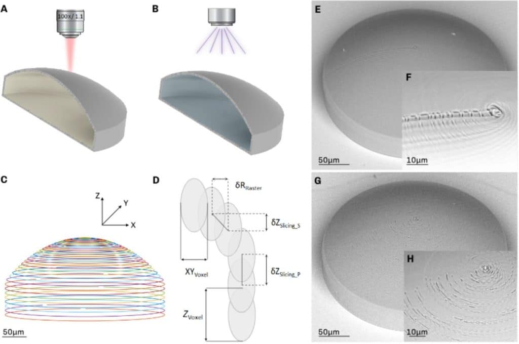
Microlenses fabricated by two-photon laser polymerization for intravital cell imaging with non-linear excitation microscopy
M. Marini, A. Nardini, R. Martínez Vázquez, C. Conci, M. Bouzin, M. Collini, R. Osellame, G. Cerullo, B.S. Kariman, M. Farsari, E. Kabouraki, M.T. Raimondi, G. Chirico
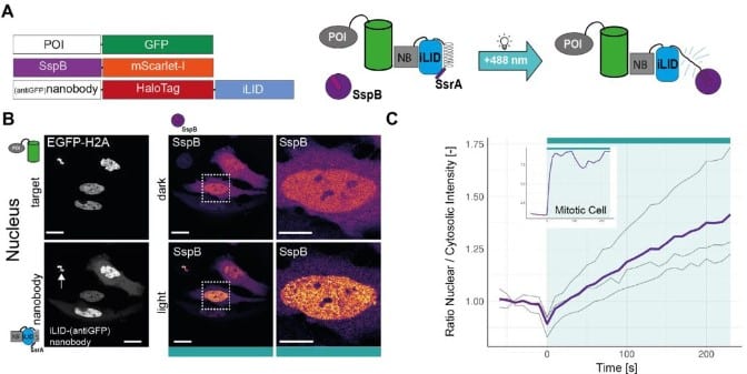
iLID-antiGFP-nanobody is a flexible targeting strategy for recruitment to GFP-tagged proteins
ProfileEike K. Mahlandt, Maarten Toereppel, Tayeba Haydary, ProfileJoachim Goedhart
 (1 votes, average: 1.00 out of 1)
Get involved
Create an account or log in to post your story on FocalPlane.
More posts like this

Filter by
- NewsApply
- DiscussionsApply
- How toApply
- ToolsApply
- Case studiesApply
- InterviewsApply
- JobsApply
- EducationApply
- Blog seriesApply
- WAMBIAN: West Africa.. in FocusApply
- Volume EMApply
- Latin American Micro..scopistsApply
- Bio-image Analysis w..ith NapariApply
- Imaging with…Apply
- Towards Global Acces..sApply
- Latin America Bioima..gingApply
- From Zero to Qupath ..HeroApply
- Asian Microscopists ..and Cell BiologistsApply
- AIC at HHMI JaneliaApply
- Deep Learning for Bi..o-image analysisApply
- GloBIAS – updates fr..om the communityApply
- Highlights from Euro..-BioImagingApply
- LSFM seriesApply
- DIY MicroscopyApply
- View all