Microscopy preprints – new tool and techniques in imaging
Posted by FocalPlane, on 14 March 2023
Here is a curated selection of preprints posted recently on new tools and techniques in imaging. Let us know if we are missing any preprints that are on your reading list!
Smart Lattice Light Sheet Microscopy for imaging rare and complex cellular events
Yu Shi, Jimmy S. Tabet, Daniel E. Milkie, Timothy A. Daugird, Chelsea Q. Yang, Andrea Giovannucci, Wesley R. Legant
Standing wave mesoscopy
Shannan Foylan, Jana Katharina Schniete, Lisa Sophie Kölln, John Dempster, Carsten Gram Hansen, Michael Shaw, Trevor John Bushell, Gail McConnell
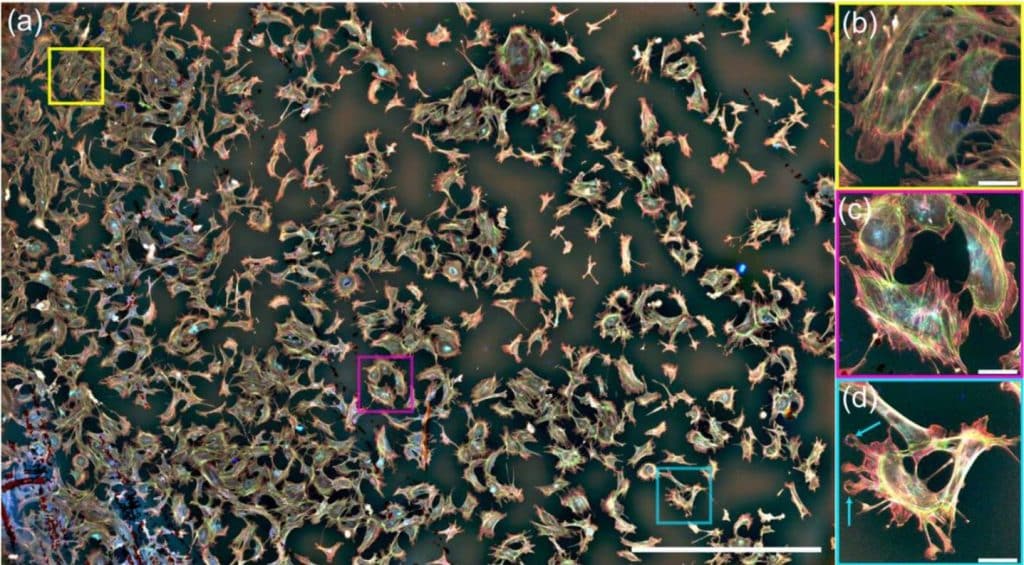
Visualizing proteins by expansion microscopy
Ali H. Shaib, Abed Alrahman Chouaib, Rajdeep Chowdhury, Daniel Mihaylov, Chi Zhang, Vanessa Imani, Svilen Veselinov Georgiev, Nikolaos Mougios, Mehar Monga, Sofiia Reshetniak, Tiago Mimoso, Han Chen, Parisa Fatehbasharzad, Dagmar Crzan, Nadia Alawar, Janna Eilts, Kim Ann Saal, Jinyoung Kang, Luis Alvarez, Claudia Trenkwalder, Brit Mollenhauer, Tiago F. Outeiro, Sarah Koester, Julia Preobraschenski, Ute Becherer, Tobias Moser, Edward S. Boyden, A. Radu A. Aricescu, Markus Sauer, Felipe Opazo, Silvio Rizzoli
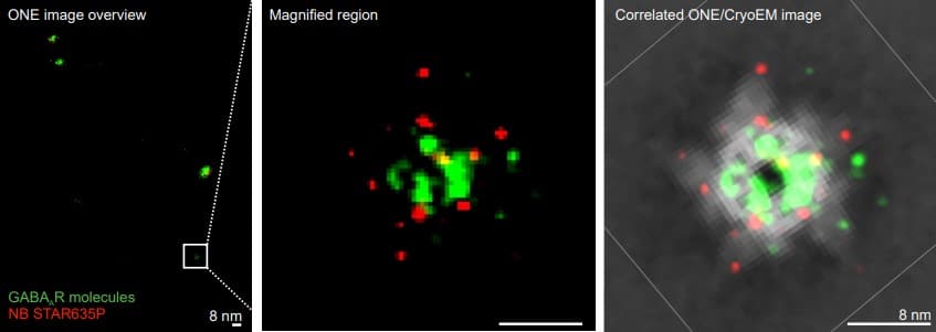
Super-resolution fluorescence imaging of cryosamples does not limit achievable resolution in cryoEM
Mart G. F. Last, Willem E.M. Noteborn, Lenard M. Voortman, Thomas H. Sharp
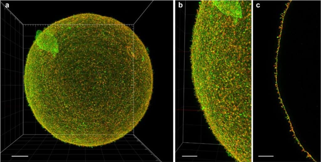
A sample preparation procedure enables acquisition of 2-channel super-resolution 3D STED image of an entire oocyte
Michaela Frolikova, Michaela Blazikova, Martin Capek, Helena Chmelova, Jan Valecka, Veronika Kolackova, Eliska Valaskova, Martin Gregor, Katerina Komrskova, Ondrej Horvath, Ivan Novotny
Shortwave-Infrared Line-Scan Confocal Microscope for Deep Tissue Imaging in Intact Organs
Jakob G. P. Lingg, Thomas S. Bischof, Bernardo A. Arús, Emily D. Cosco, Ellen M. Sletten, Christopher J. Rowlands, Oliver T. Bruns, Andriy Chmyrov
STORM imaging buffer with refractive index matched to standard immersion oil
Youngseop Lee, Yeunho Lee, Minchol Lee, Donghoon Koo, Dongwoo Kim, Kangwon Lee, Jeongmin Kim
When Optical Microscopy Meets All-Optical Analog Computing: A Brief Review
Yichang Shou, Jiawei Liu, Hailu Luo
T-CLEARE: A Pilot Community-Driven Tissue-Clearing Protocol Repository
Kurt Weiss, Jan Huisken, Vesselina Bakalov, Michelle Engle, Lauren Gridley, Michelle Krzyzanowski, Tom Madden, Deborah Maiese, Justin Waterfield, David Williams, Xin Wu, Carol Marie Hamilton, Wayne Huggins
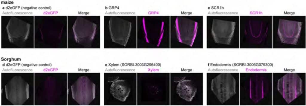
A rapid and sensitive multiplex, whole mount RNA fluorescence in situ hybridization and immunohistochemistry protocol
Tian Huang, Bruno Guillotin, Ramin Rahni, Ken Birnbaum, Doris Wagner
Selection of antibody-binding covalent aptamers
Noah Soxpollard, Sebastian Strauss, Ralf Jungmann, Iain MacPherson
An expanded GCaMP reporter toolkit for functional imaging in C. elegans
Jimmy Ding, Lucinda Peng, Sihoon Moon, Hyun Jee Lee, Dhaval S. Patel, Hang Lu
Mid-infrared Chemical Imaging of Intracellular Tau Fibrils using Fluorescence-guided Computational Photothermal Microscopy
Jian Zhao, Lulu Jiang, Alex Matlock, Yihong Xu, Jiabei Zhu, Hongbo Zhu, Lei Tian, Benjamin Wolozin, Ji-Xin Cheng
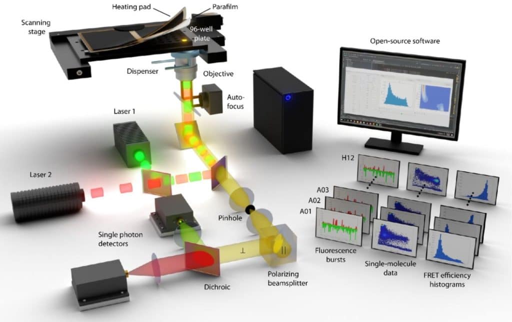
Farewell to single-well: An automated single-molecule FRET platform for high-content, multiwell plate screening of biomolecular conformations and dynamics
Andreas Hartmann, Koushik Sreenivasa, Mathias Schenkel, Neharika Chamachi, Philipp Schake, Georg Krainer, Michael Schlierf
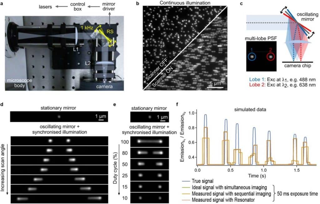
Fast and artifact-free excitation multiplexing using synchronized image scanning
Ezra Bruggeman, Robin Van den Eynde, Baptiste Amouroux, Tom Venneman, Pieter Vanden Berghe, Marcel Müller, Wim Vandenberg, Peter Dedecker
Assessing the performance of the Cell Painting assay across different imaging systems
Nasim Jamali, Callum Tromans-Coia, Hamdah Shafqat Abbasi, Kenneth A. Giuliano, Mai Hagimoto, Kevin Jan, Erika Kaneko, Stefan Letzsch, Alexander Schreiner, Jonathan Z. Sexton, Mahomi Suzuki, O. Joseph Trask, Mitsunari Yamaguchi, Fumiki Yanagawa, Michael Yang, Anne E. Carpenter, Beth A. Cimini
Geometry-preserving Expansion Microscopy microplates enable high fidelity nanoscale distortion mapping
Rajpinder S. Seehra, Benjamin H.K. Allouis, Thomas M.D. Sheard, Michael E Spencer, Tayla Shakespeare, Ashley Cadby, Izzy Jayasinghe
A statistical resolution measure of fluorescence microscopy with finite photons
Yilun Li, Fang Huang
Optimization of highly inclined Illumination for diffraction-limited and super-resolution microscopy
L. Gardini, T. Vignolini, V. Curcio, F.S. Pavone, M. Capitanio
POLCAM: Instant molecular orientation microscopy for the life sciences
Ezra Bruggeman, Oumeng Zhang, Lisa-Maria Needham, Markus Körbel, Sam Daly, Matthew Cheetham, Ruby Peters, Tingting Wu, Andrey S. Klymchenko, Simon J. Davis, Ewa K. Paluch, David Klenerman, Matthew D. Lew, Kevin O’Holleran, Steven F. Lee
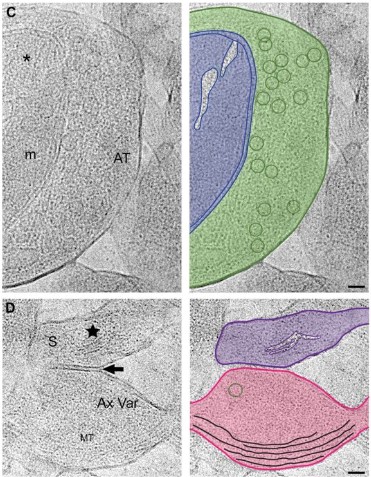
Cryo-FIB workflow for imaging brain tissue via in situ cryo-electron microscopy
Jiying Ning, Jill R. Glausier, Chyongere Hsieh, Thomas Schmelzer, Silas A. Buck, Jonathan Franks, Cheri M. Hampton, David A. Lewis, Michael Marko, Zachary Freyberg
A fluorogenic chemically induced dimerization technology for controlling, imaging and sensing protein proximity
Sara Bottone, Zeyneb Vildan Cakil, Octave Joliot, Gaelle Boncompain, Franck Perez, Arnaud Gautier
Optofluidic adaptive optics in multi-photon microscopy
Maximilian Sohmen, Juan D. Muñoz-Bolaños, Pouya Rajaeipour, Monika Ritsch-Marte, Çağlar Ataman, Alexander Jesacher
Boosting the toolbox for live imaging of translation
Maelle Bellec, Ruoyu Chen, Jana Dhayni, Cyril Favard, Antonello Trullo, Helene Lenden-Hasse, Ruth Lehmann, Edouard Bertrand, Mounia Lagha, Jeremy Dufourt
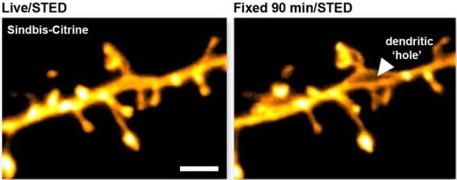
The impact of chemical fixation on the microanatomy of mouse brain tissue
Agata Idziak, V.V.G. Krishna Inavalli, Stephane Bancelin, Misa Arizono, U. Valentin Nägerl
Differentiable Microscopy for Content and Task Aware Compressive Fluorescence Imaging
Udith Haputhanthri, Andrew Seeber, Dushan Wadduwage
 (No Ratings Yet)
2 thoughts on “Microscopy preprints – new tool and techniques in imaging”
Leave a Reply
Get involved
Create an account or log in to post your story on FocalPlane.
More posts like this

Filter by
- NewsApply
- DiscussionsApply
- How toApply
- ToolsApply
- Case studiesApply
- InterviewsApply
- JobsApply
- EducationApply
- Blog seriesApply
- Volume EMApply
- Latin American Micro..scopistsApply
- Bio-image Analysis w..ith NapariApply
- Imaging with…Apply
- Towards Global Acces..sApply
- Latin America Bioima..gingApply
- From Zero to Qupath ..HeroApply
- Asian Microscopists ..and Cell BiologistsApply
- AIC at HHMI JaneliaApply
- Deep Learning for Bi..o-image analysisApply
- GloBIAS – updates fr..om the communityApply
- Highlights from Euro..-BioImagingApply
- LSFM seriesApply
- DIY MicroscopyApply
- View all
Dear FocalPlane team, since you ask in your newsletter, I`m actually missing this cool pre-print: https://www.biorxiv.org/content/10.1101/2022.10.06.511114v1
Thanks Maria, definitely a very impressive piece of work. We included the preprint in our October list: https://focalplane.biologists.com/2022/10/21/microscopy-preprints-new-tool-and-techniques-in-imaging-2/
Do let us know about any other preprints that you are enjoying!