Featured image with Ellen Skarpen and José Teles Reis
Posted by FocalPlane, on 5 April 2023
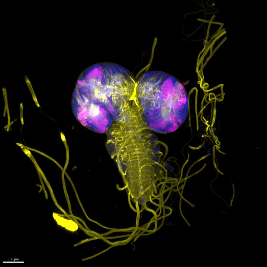
Our featured image shows a Drosophila third instar larval brain with activated ‘Mosaic analysis with a repressible cell marker’ (MARCM) specifically in the Optic lobe, marked by GFP. This genetic technique allows for the labelling and manipulation of mitotically active clones of cells for the study of fate mapping or gene role through gain/loss of function. In this image we can clearly observe the neuropil of the Drosophila larval brain and the segmental nerves leaving the ventral nerve chord to innervate the peripheral organs. This sample was microdissected from the larva, fixed in PFA and later stained for DNA (Hoechst) and the actin cytoskeleton (Phalloidin 568). GFP is expressed genetically in MARCM activated clones through the UAS-GAL4 system. The sample was imaged on a Nikon spinning disc with a 25X SIL Objective with optical sectioning covering the whole organ. Visualization in 3D-view was performed using Imaris, where we chose to present Hoechst in blue, GFP in magenta, and Phalloidin in yellow.
The sample was prepared by José Teles Reis, and imaged by Ellen Skarpen.
José Teles Reis is a PhD student in the lab of Tor Erik Rusten at the Institute for Cancer Research in Oslo, Norway. Previously, he was in the lab of Catarina Homem at NOVA Medical School, Lisbon, Portugal, where he worked on characterising neuroblast lineage development at the single-cell level. Currently, José is designing novel genetic systems for the study of tumour-host interactions and is interested in the molecular mechanisms behind tumour-microenvironment crosstalk. José’s favourite imaging technique is spinning disk microscopy, which allows multi-channel fast acquisition of thick samples for 3D reconstruction and the study of collective cell behaviour in Drosophila tissues.
Ellen Skarpen is the unit leader of the Advanced Microscopy facility at the Institute for Cancer Research in Oslo, Norway. Her research background is within cellular signaling pathways, toxicology, and cancer research. Currently, she supports research projects within membrane dynamics and its role in cancer-related processes like proliferation, migration, and invasion. Ellen’s favourite imaging technique is confocal spinning disk microscopy due its combination of gentle imaging, high speed, and high resolution in 2D- and 3D. Ellen is excited by future possibilities of advanced image analysis.
 (4 votes, average: 1.00 out of 1)