Microscopy preprints – new tools and techniques in imaging
Posted by FocalPlane, on 16 June 2023
Here is a curated selection of preprints posted recently on new tools and techniques in imaging. Let us know if we are missing any preprints that are on your reading list!
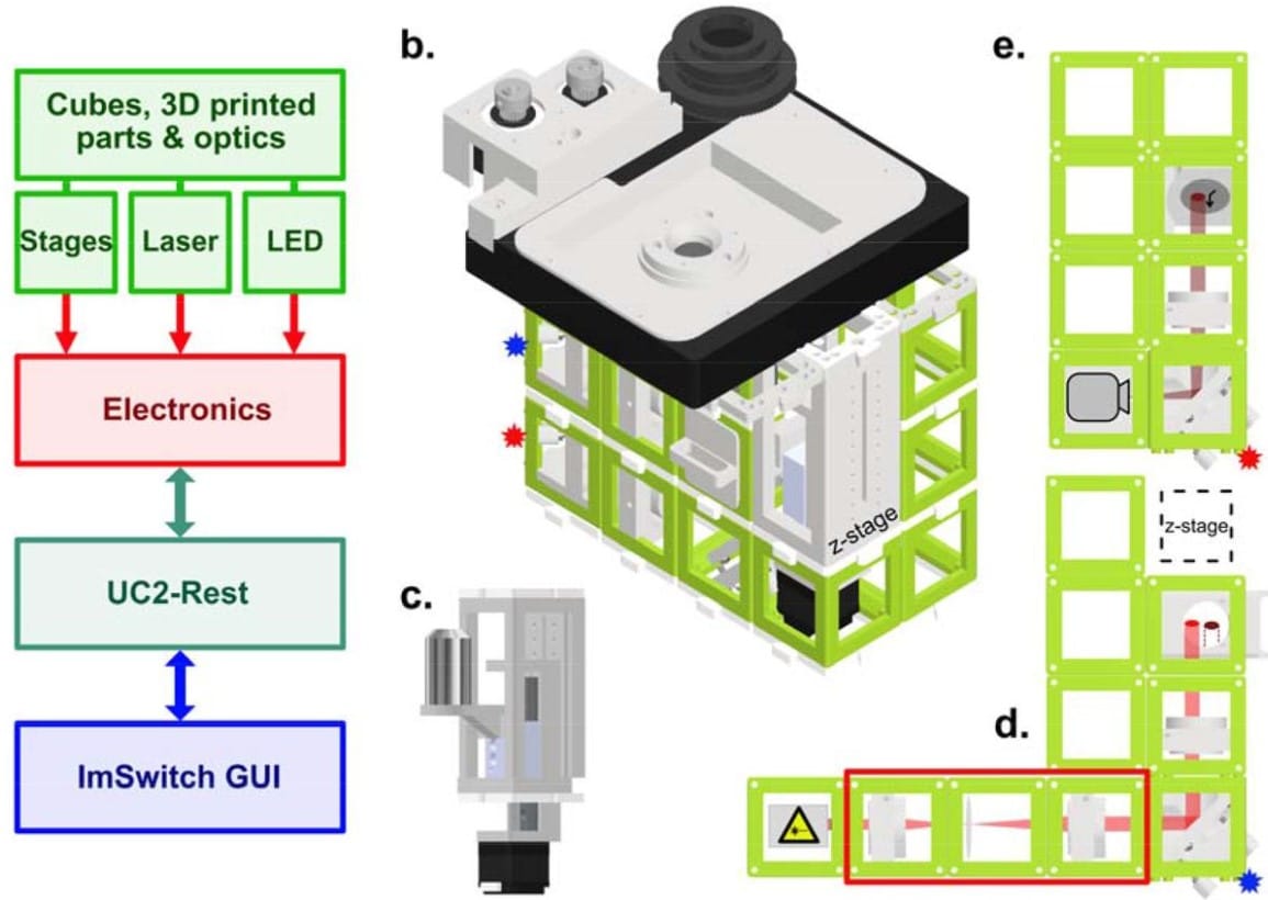
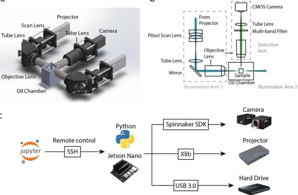
An open-source, high resolution, automated fluorescence microscope
Ando C. Zehrer, Ana Martin-Villalba, Benedict Diederich, Helge Ewers
Signal Improved ultra-Fast Light-sheet Microscope (SIFT) for large tissue imaging
Md Nasful Huda Prince, Benjamin Garcia, Cory Henn, Yating Yi, Etsuo A. Susaki, Yuki Watakabe, Tomomi Nemoto, Keith A Lidke, Hu Zhao, Irene Salinas Remiro, Sheng Liu, Tonmoy Chakraborty

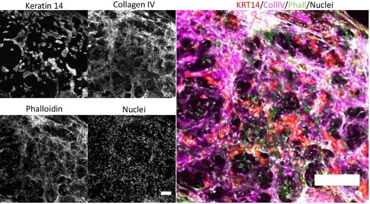
Streamlined intravital imaging approach for long-term monitoring of epithelial tissue dynamics on an inverted confocal microscope
Michael Hamersky IV, Khushi Tekale, L. Matthew Winfree, Matthew JM Rowan, Lindsey Seldin
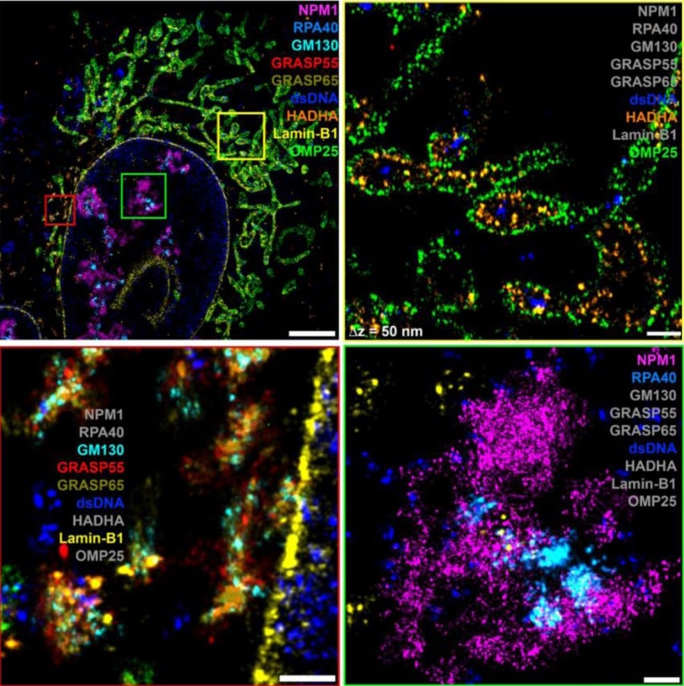
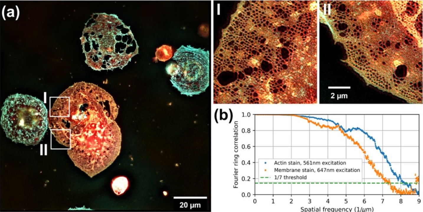
Unraveling cellular complexity with unlimited multiplexed super-resolution imaging
Florian Schueder, Felix Rivera-Molina, Maohan Su, Phylicia Kidd, James E. Rothman, Derek Toomre, Joerg Bewersdorf
Expansion-assisted selective plane illumination microscopy for nanoscale imaging of centimeter-scale tissues
Adam Glaser, Jayaram Chandrashekar, Joshua Vasquez, Cameron Arshadi, Naveen Ouellette, Xiaoyun Jiang, Judith Baka, Gabor Kovacs, Micah Woodard, Sharmishtaa Seshamani, Kevin Cao, Nathan Clack, Andrew Recknagel, Anna Grim, Pooja Balaram, Emily Turschak, Alan Liddell, John Rohde, Ayana Hellevik, Kevin Takasaki, Lindsey Erion Barner, Molly Logsdon, Chris Chronopoulos, Saskia de Vries, Jonathan Ting, Steve Perlmutter, Brian Kalmbach, Nikolai Dembrow, R. Clay Reid, David Feng, Karel Svoboda
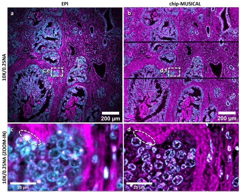
Super-resolution histology of paraffin-embedded samples via photonic chip-based microscopy
Luis E. Villegas-Hernández, Vishesh K. Dubey, Hong Mao, Manohar Pradhan, Jean-Claude Tinguely, Daniel H. Hansen, Sebastian Acuña, Bartłomiej Zapotoczny, Krishna Agarwal, Mona Nystad, Ganesh Acharya, Kristin A. Fenton, Håvard E. Danielsen, Balpreet S. Ahluwalia
High-throughput confocal airy beam oblique light-sheet tomography of brain-wide imaging at single-cell resolution
Xiaoli Qi, Rodrigo Muñoz-Castañeda, Arun Narasimhan, Liya Ding, Xin Chen, Corey Elowsky, Jason Palmer, Rhonda Drewes, Jianjun Sun, Judith Mizrachi, Hanchuan Peng, Zhuhao Wu, Pavel Osten
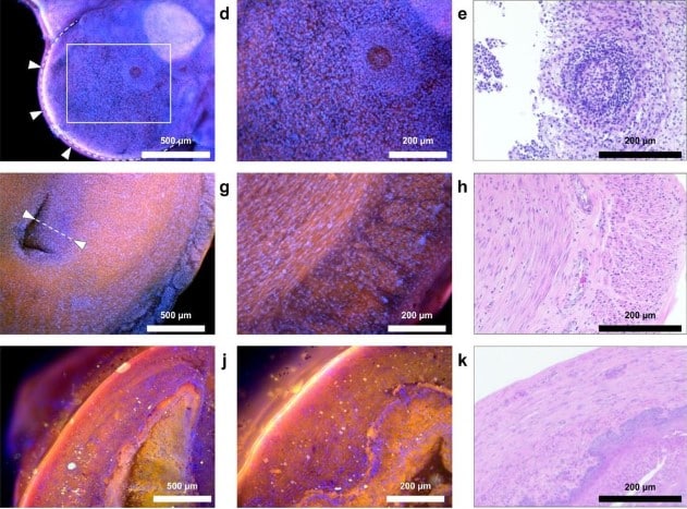
Deep ultraviolet-excited fluorescence histology with fibre optic illumination for tissue microtopography and quantitative mapping
Alexander Si Kai Yong, Ko Hui Tan, Joel Lang Yi Ang, Chiyo Wan Xuan Tan, Jessica Sze Jia Kng, Cyrus Jia Jun Tan, Rachael Hui Kie Soh, Kaicheng Liang
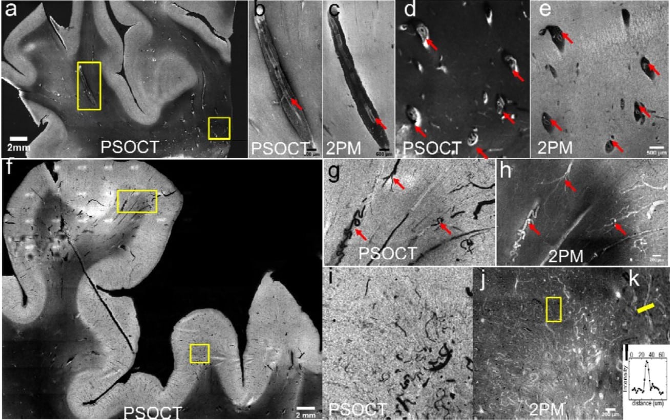
Multi-Scale Label-free Human Brain Imaging with Integrated Serial Sectioning Polarization Sensitive Optical Coherence Tomography and Two-Photon Microscopy
Shuaibin Chang, Jiarui Yang, Anna Novoseltseva, Xinlei Fu, Chenglin Li, Shih-Chi Chen, Jean C. Augustinack, Caroline Magnain, Bruce Fischl, Ann C. Mckee, David A. Boas, Ichun Anderson Chen, Hui Wang
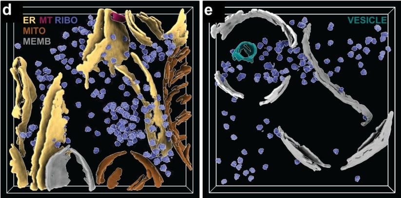
Serialized On-grid Lift-In Sectioning for Tomography (SOLIST)
Nguyen Ho Thuy Dung, Gaia Perone, Roberta Vazzana, Flaminia Kaluthantrige Don, Malan Silva, Simona Sorrentino, Paolo Swuec, Frederic Leroux, Nereo Kalebic, Francesca Coscia, Philipp S. Erdmann
High-speed TIRF and 2D super-resolution structured illumination microscopy with large field of view based on fiber optic components
Henning Ortkrass, Jasmin Schürstedt, Gerd Wiebusch, Karolina Szafranska, Peter Mccourt, Thomas Huser
High-efficiency digitally scanned light-sheet fluorescence lifetime microscopy (DSLM-FLIM)
Kyle J. Nutt, Daniel Olesker, Ewan McGhee, Graham Hungerford, Christopher G. Leburn, Jonathan Taylor
Metabolic FRET sensors in intact organs: Applying spectral unmixing to acquire reliable signals
Lautaro Gándara, Lucía Durrieu, Pablo Wappner
FluoMALDI microscopy: matrix co-crystallization simultaneously enhances fluorescence and MALDI imaging
Ethan Yang, Xinyi Elaine Shen, Hoku West-Foyle, Dalton R. Brown, Cole C. Johnson, Jeong Hee Kim, LaToya Ann Roker, Caitlin M. Tressler, Ishan Barman, Scot C. Kuo, Kristine Glunde
Laser patterning bioprinting using a light sheet-based system equipped with light sheet imaging produces long-term viable skin constructs
Levin Hafa, Louise Breideband, Lucas Ramirez Posada, Núria Torras, Elena Martinez, Ernst H.K. Stelzer, Francesco Pampaloni
Scalable projected Light Sheet Microscopy for high-resolution imaging of large samples
Yannan Chen, Cheng Gong, Shradha Chauhan, Estanislao Daniel De La Cruz, Malika S. Datta, Raju Tomer
Indirect Correlative Light and Electron Microscopy (iCLEM): A Novel Pipeline for Multiscale Quantification of Structure from Molecules to Organs
Heather L. Struckman, Nicolae Moise, Bieke Vanslembrouck, Nathan Rothacker, Zhenhui Chen, Jolanda van Hengel, Seth H. Weinberg, Rengasayee Veeraraghavan
Proximal Molecular Probe Transfer (PROMPT), a new approach for identifying sites of protein/nucleic acid interaction in cells by correlated light and electron microscopy
Guillaume A Castillon, Sebastien Phan, Junru Hu, Daniela Boassa, Stephen R Adams, Mark H Ellisman
A 3D-printed flow-cell for on-grid purification of electron microscopy samples directly from lysate
Kailash Ramlaul, Ziyi Feng, Caoimhe Canavan, Martín Natàlia de Garrido, David Carreño, Michael Crone, Kirsten E. Jensen, Bing Li, Harry Barnet, David T. Riglar, Paul S. Freemont, David Miller, Christopher H. S. Aylett
Long-term optical imaging of the spinal cord in awake, behaving animals
Biafra Ahanonu, Andrew Crowther, Artur Kania, Mariela Rosa Casillas, Allan Basbaum

In-section Click-iT detection and super-resolution CLEM: Shedding light on nucleolar ultrastructure and S-phase progression in plants
M Franek, L Koptasikova, J Miksatko, J Pospisil, D Liebl, M Esner, E Macickova, M Dvorackova, J Fajkus

Post-acquisition super resolution for cryo-electron microscopy
Raymond N. Burton-Smith, Kazuyoshi Murata
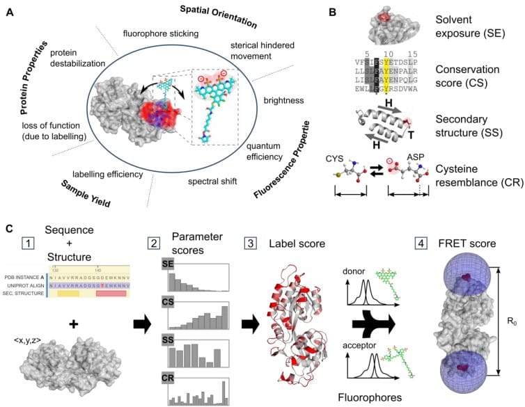
Labelizer: systematic selection of protein residues for covalent fluorophore labeling
Christian Gebhardt, Pascal Bawidamann, Konstantin Schuetze, Gabriel Gustavo Moya Munoz, Anna-Katharina Spring, Douglas Griffith, Jan Lipfert, Thorben Cordes
Multiplexed volumetric CLEM enabled by antibody derivatives provides new insights into the cytology of the mouse cerebellar cortex
Xiaomeng Han, Xiaotang Lu, Peter H. Li, Shuohong Wang, Richard Schalek, Yaron Meirovitch, Zudi Lin, Jason Adhinarta, Daniel Berger, Yuelong Wu, Tao Fang, Elif Sevde Meral, Shadnan Asraf, Hidde Ploegh, Hanspeter Pfister, Donglai Wei, Viren Jain, James S. Trimmer, Jeff W. Lichtman
Spatial proteomics in neurons at single-protein resolution
Eduard M. Unterauer, Sayedali Shetab Boushehri, Kristina Jevdokimenko, Luciano A. Masullo, Mahipal Ganji, Shama Sograte-Idrissi, Rafal Kowalewski, Sebastian Strauss, Susanne C.M. Reinhardt, Ana Perovic, Carsten Marr, Felipe Opazo, Eugenio F. Fornasiero, Ralf Jungmann

 (No Ratings Yet)
Get involved
Create an account or log in to post your story on FocalPlane.
More posts like this

Filter by
- NewsApply
- DiscussionsApply
- How toApply
- ToolsApply
- Case studiesApply
- InterviewsApply
- JobsApply
- EducationApply
- Blog seriesApply
- Volume EMApply
- Latin American Micro..scopistsApply
- Bio-image Analysis w..ith NapariApply
- Imaging with…Apply
- Towards Global Acces..sApply
- Latin America Bioima..gingApply
- From Zero to Qupath ..HeroApply
- Asian Microscopists ..and Cell BiologistsApply
- AIC at HHMI JaneliaApply
- Deep Learning for Bi..o-image analysisApply
- GloBIAS – updates fr..om the communityApply
- WAMBIAN: West Africa.. in FocusApply
- Highlights from Euro..-BioImagingApply
- LSFM seriesApply
- DIY MicroscopyApply
- View all