Featured image with Martín Estermann
Posted by FocalPlane, on 15 September 2023
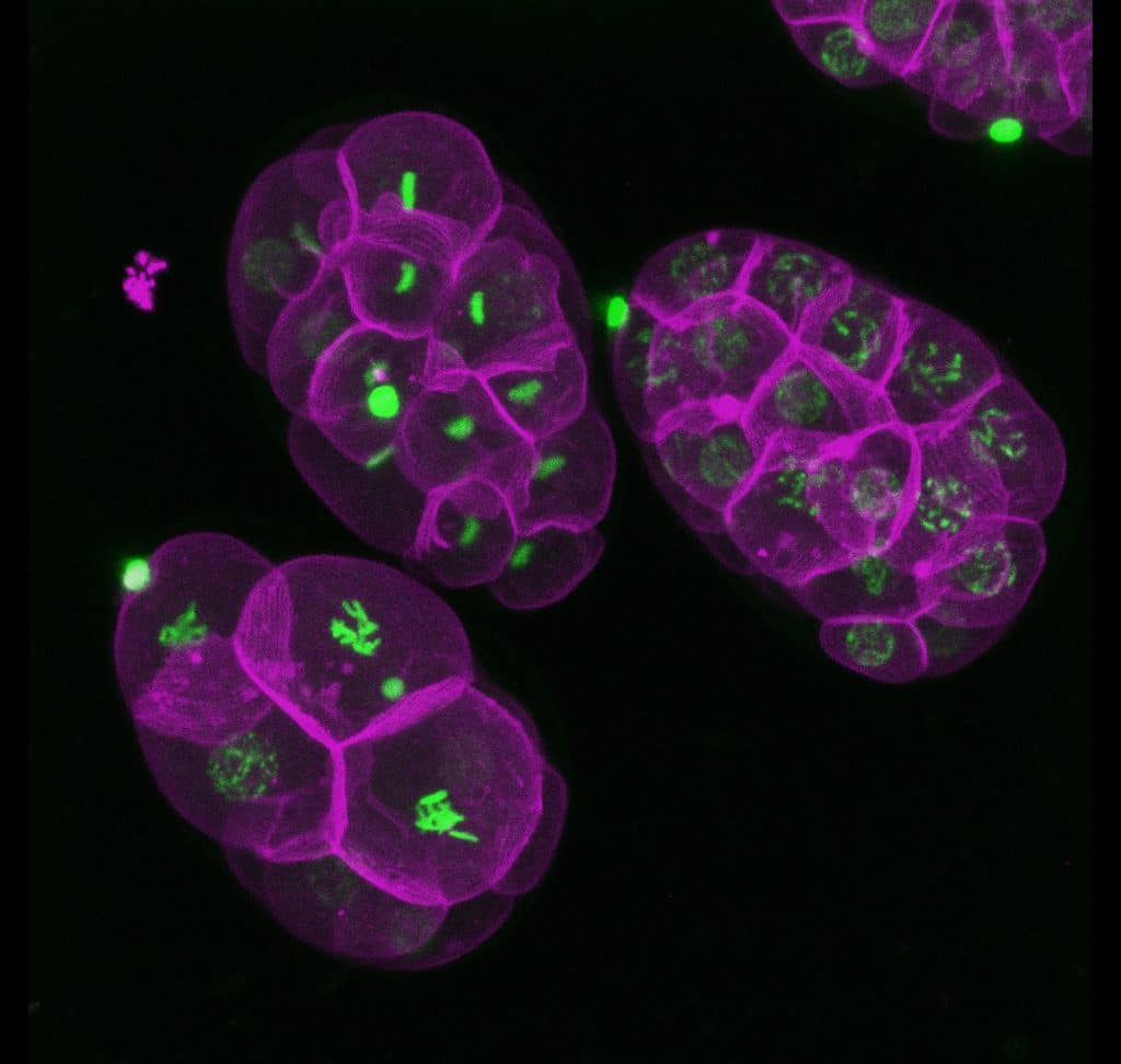
Our featured image, acquired by Martín Estermann, is a 3D reconstruction of C. elegans transgenic embryos. Embryos were imaged live using a Zeiss Celldiscoverer 7 microscope. The 3D reconstruction was performed using Fiji. Histones in are shown in green and the cell membrane in magenta.
We caught up with Martín to find out about his research and what he is excited about in microscopy.
Research career so far: I did my undergrad in Argentina at UNNOBA university. Then I moved to Melbourne, Australia to do my PhD at Monash University. Currently, I am a postdoctoral fellow at NIEHS, North Carolina, USA.
Current research: I am particularly interested in genetics, developmental biology, evo-devo, and specifically in sex determination and gonadal development.
Favourite imaging technique/microscope: I love fluorescence microscopy. I mostly work with confocal microscopes, but I am trying to get more training using light-sheet microscopy.
What are you most excited about in microscopy? I am really interested in visualizing embryonic development in real time using microscopy. I want to move from the 2D imaging of histological sections to the 3D imaging of whole organs or embryos. I am really attracted to 3D renderings of fluorescently labelled embryos from different species at different developmental time points; they are mesmerizing.
Martín is an Ambassador for our sister site, preLights. You can read their interview with Martín here: https://prelights.biologists.com/news/meet-the-prelights-ambassadors-martin-estermann/
 (No Ratings Yet)