Featured image with Ioakeim (Makis) Ampartzidis
Posted by FocalPlane, on 27 October 2023
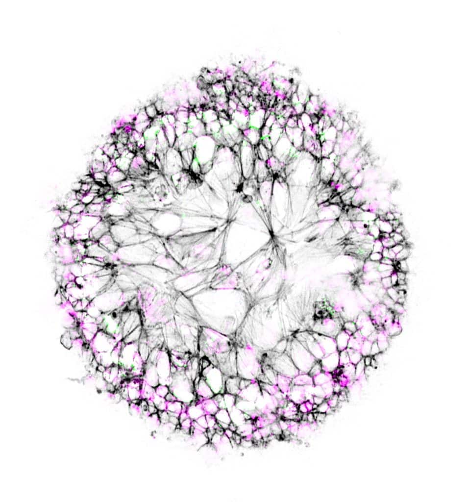
Our featured image, ‘Dandelion in spring’ from Ioakeim (Makis) Ampartzidis, shows neuroepithelial-like cells cultured on glass beads. The cell nuclei are coloured in magenta, and the cytoskeleton is stained in black, labelling F-actin. The actively proliferating cells are highlighted with Ki-67 antigen in green.
We caught up with Makis to find out more about his research and what he is excited about in microscopy.
Research career so far: I completed my undergraduate studies in the Department of Molecular Biology and Genetic at the Democritus University of Thrace, Greece in 2019. A year later, I completed my MPhil studies in the Boroviak lab at University of Cambridge, before starting my PhD at UCL in 2020. Currently, I am a final-year PhD student in the Galea lab based at the Great Ormond Street Institute of Child Health, London. In the neurulation biomechanics lab, we study neural tube defects using animal models, patient-derived tissue and human induced-pluripotent stem cells.
Current research: My research focusses on disease modelling and regenerative strategies for spina bifida. During my PhD I have developed and characterised a neuroepithelial platform using human induced-pluripotent cells (hiPSCs), in which I validated various bona-fide neuroepithelial behaviours in vitro, including apical constriction. To better understand neuroepithelial-specific pathological mechanisms, I use hiPSCs that are engineered to carry patient-identified genetic alterations, as well as patient-derived material. My research aims to elucidate the cellular mechanisms that underlie the development of the spina bifida.
Favourite imaging technique/microscope: Throughout my PhD, I have used the Zeiss LSM 880 confocal to better characterise the neuroepithelial system. The versatility of the system to obtain high resolution images through Airyscan detection set up, and to perform laser ablation in live cells was insightful for my project.
What are you most excited about in microscopy? Recently, I have started exploring widefield microscopy, primarily for live imaging of labelled cells. For post-acquisition image processing I use deconvolution software which is essential for increasing the signal-to-noise ratio and resolution of my images. In the future, I look forward to exploring other imaging systems, such as light-sheet and spinning-disk confocal microscopes.
 (2 votes, average: 1.00 out of 1)