Crick Bioimage Analysis Symposium 2023 – a Review
Posted by vanesssadao, on 22 December 2023
(By Vanessa Dao, Hradini Konthalapalli, Olatz Niembro Vivanco, Karishma Valand)
The Crick Bioimage Analysis Symposium had its first in-person meeting in 2022. This year, #CBIAS2023 gathered around 200 people on site and 80 virtually. It has been an exciting two days of bringing biomedical researchers and their questions together with image analysis and their techniques. Are you wondering what that might look like? Join us –Olatz, an immunology PhD student, Hradini, a cell biology PhD student, Karishma, a cancer biology PhD student and Vanessa, a physics undergraduate – as we share our experience. Maybe next year you will join us at The Crick too!
Program Structure
(Karishma) This year CBIAS was held in a hybrid format, which was beneficial for attendees who could not join in person but wanted to attend the talks. Invited speakers had expertise ranging from developing imaging modalities to computational tools to biological implications. The schedule allowed the talks to flow nicely into one another. The talks ranged from 5 to 30 minutes and were arranged in a way that did not make the audience feel overwhelmed by information. There were also ample opportunities for networking during the coffee breaks, lunch and the evening social. It was great to meet and interact with so many people from different areas of expertise coming together and sharing their knowledge on image analysis. One critique here was maybe the lunch session was a bit too long (1 hr 45 mins) and could have been shorter to allow for more questions after each speaker. Overall, it was a well organised event that brought together a wide range of speakers and attendees.
Poster Session
You can navigate over 30 posters (including Vanessa’s #1!) from the symposium by following this link.

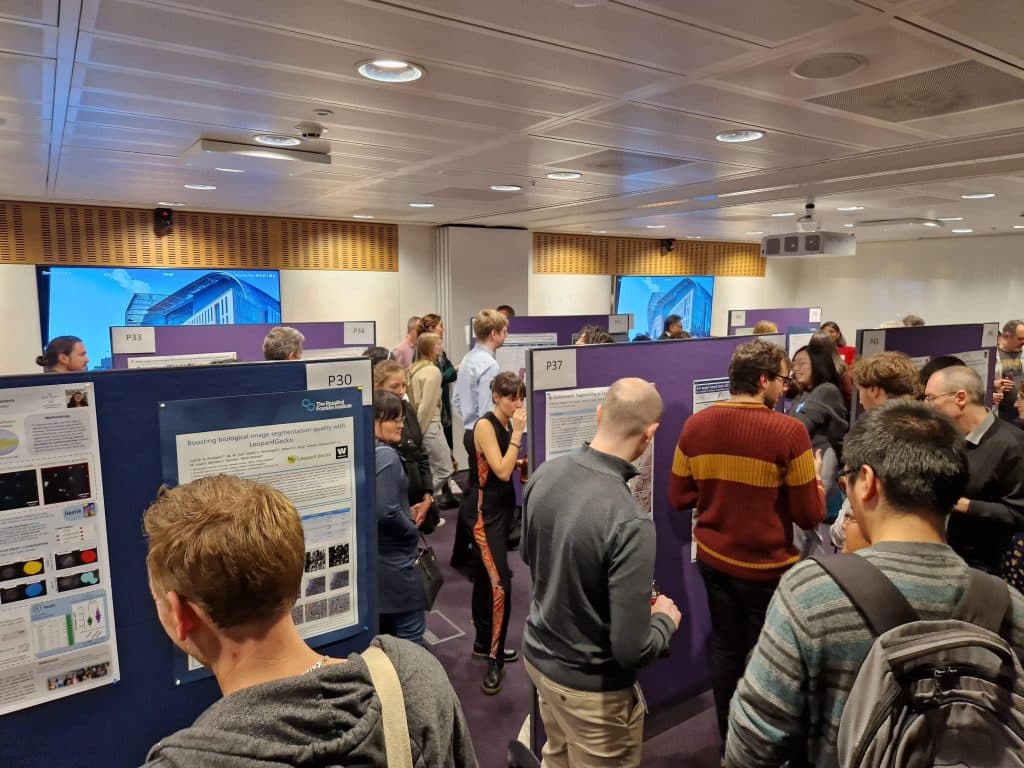
(Vanessa) The poster session had 55 high-quality diverse posters presented which fostered many conversations around new computational tools and user experiences. This was my first time presenting a poster and I found it to be a very rewarding experience – not just because I had the honour of winning Best Poster – but because I gained lots of valuable feedback on how I could develop my project further. These networking events provide an amazing opportunity to share your work and invite discussions from various perspectives which make for great scientific exchange in a welcoming environment. It was a very inspiring and insightful experience to see the community effort in making bioimage analysis easier for everyone.
(Olatz) The poster session was held over drinks and nibbles, and even though the space was somewhat crowded and loud, it was easy to weave around and spot posters that spark your interest. The topics covered everything from the very state of the art of tool development to all the exciting ways biology is implementing them, discussions flowing nicely between the two. The diverse and welcoming atmosphere, much like throughout the entire conference, made it a great networking opportunity.
Discussion Panel Highlights
If you want to immerse in the discussions yourself and find out which panelist actively tried to start a fight, both panel sessions as well as most of the talks are now available on youtube here.
(O) The first day closed with an exciting panel discussion. Chair Marie Held (University of Liverpool) was accompanied by Christian Tischer (EMBL Heidelberg), Stefania Marcotti (King’s College London), and Tatiana Woller (VIB). Together, they delved into the evolving role of bioimage analysts within institutions, as NEUBIAS grows into GLOBIAS, and the emphasis shifts towards quantitative bioimaging over qualitative work. A vibrant part of the discussion revolved around the challenge faced by local experts who have inadvertently assumed the role of informal image analysts without formal recognition or compensation. Many analysts currently find themselves in awkward or non-existent institutional positions, but through these discussions, initiatives like CBIAS are laying the groundwork for a supportive platform. Not that long ago NEUBIAS coined the title of Bioimage Analyst, contributing to the community’s growth and its ability to advocate for designated positions within institutions. The panel shared success stories, such as the establishment of the Research Technical Professional Career pathway at the University of Liverpool. When bioimage analysts are situated in organised roles within and across institutions, the panel argued there is a greater chance of implementing structured and standardised user training, which has the potential to raise the standards of quantitative bioimaging, starting from even before image acquisition. And as the experimentalists’ understanding of what is possible increases, so will our ability to address both longstanding and novel biological questions
(V) Big questions involving the future of bioimage analysis were asked on the second panel of the meeting, chaired by Martin Jones (Francis Crick) and answered by Beth Cimini (Broad Institute), Aybuke Kupcu Yoldas (EMBL-EBI), Norman Rzepka (Scaleable Minds) and Kimberly Meechan (UCL). It is safe to say that no one can predict what will happen within this community, bioimage analysis is a fast-evolving field with new tools and methods being developed constantly. It was highlighted by Beth that image.sc is only 5 years old and Cellpose only 3 – how could we possibly imagine what will be achieved in 1, 5 years, let alone 10! With this in mind, the emphasis on accessibility has paved the way for NGFFs and web architectures such as Webknossos and Piximi to support groups – who do not have the infrastructure required – with accessing and analysing data on the fly without the need to download anything. Hopefully, this will lead to continual growth in the community, promoting the sharing of data and analyses. Additionally, it was pointed out that microscope manufacturers should be included in ongoing discussions to collectively establish standards for the efficient storage and transfer of image data and metadata.
Talk Highlights
(V) The influence of artificial intelligence in bioimage analysis was clearly seen throughout the symposium; many talks presented the application of AI in workflows including CLEM-Reg and Spotipy leveraging MitoNET and SpotNet, respectively. Even with widespread use within science, Sreenivas Bhattiprolu (Zeiss) took the time to make the important distinction between conventional machine learning and deep learning in image analysis, highlighting how this can impact generalisation of models to diverse datasets and time required to fine-tune models for optimal results. As these tools are being developed for bioimage analysis, grounding the data within the context of the scientific motivation allows for making informed decisions about how to process, segment and track the images as well as how to interpret the results. With this in mind, I particularly enjoyed Kristina Ulicna (Alan Turing Institute) explain the implementation of self-supervised learning for cell cycle annotation and quantifying the uncertainty of those resultant labels.
(O) CBIAS gives a taste of where the field is headed, giving you the chance to delve into the hot topics in image analysis and in the biology that drives them. But the speakers also emphasise learning: the NEUBIAS network seminars, Introduction to Bioimage Analysis by Pete Bankhead and the Bioimaging Guide presented by Beth Cimini were a central part of the talks. While not as beginner-friendly, others like Ana Stojiljkovic demoed their software during their talk, as an inspiring taster of what can be achieved. Having these new resources under my belt might have an impact on my career prospects as I am coming to the end of my PhD.
(K) Some of my favourite talks were by Pete Bankhead and Beth Cimini. Both these talks were presented with enthusiasm, had demos of the tools, were clear, engaging and jargon free – meaning people with no prior-knowledge could still easily follow. Pete presented updates of his QuPath tool and I particularly enjoyed his anecdote of using QuPath to digitise and classify family photographs – including using the deep-learning model to classify his guinea pig! Beth demonstrated Piximi.app, a web-based deep learning tool for image analysis. Piximi can be used on any device with internet – PC, laptop and even on your phone, to perform tasks such as image segmentation and classification, and easily share with collaborators. She also presented the Bioimaging guide – a curated set of resources, tips, guides for biologists starting out in the field of bioimaging.
Accessibility and Amenities
(O) Notably, the venue was entirely step-free, and so was the venue for the social. The talks were live-streamed with real-time captions readable both online and in the room—a nice touch for accessibility. The organizers, having learned from past experiences, improved the vegan options for dietary needs. If you’re considering attending and have concerns about accessibility or dietary restrictions, believe they’ve taken steps to address these aspects and responded positively to feedback.
Will CBIAS24 be relevant for you?
(V) Overall, I think CBIAS 2023 was a well-organised event, so huge credit to the organisers and the teams working behind the scenes! The symposium catered well to both new and experienced image analysts, with a range of talks and a diverse poster selection. While not entirely aimed at newcomers, it still provided valuable insight into ongoing tool developments and challenges being addressed by the community.
It was also great seeing people come together in a joint effort to increase the global presence of bioimage analysis – I think by simply attending, a valuable contribution is made to the community by gaining more awareness of the activities within the field. Hybrid symposiums like CBIAS, along with workshops, hackathons, and the rise of image analysis facilities, strengthen the community by encouraging collaboration and introducing students to image analysis and its available opportunities. As an undergraduate student, CBIAS exposed me to accessible tools and resources useful for skill development and potential career entry into the field. For anyone looking for a thorough overview and insight into bioimage analysis, it’s definitely an event worth attending.
Lastly, image analysis is not confined to the field of biology – as seen by the diverse panels and attendees. It is important to learn about the tools and share knowledge across different disciplines to promote the development of scientific methods in all fields, making the symposium worth attending including for those outside of bioimage analysis.
(Hradini) As an early-stage PhD student, attending CBIAS felt like a warm welcome into the bioimage analysis community! The conference provided a valuable opportunity to connect with fellow researchers and bioimage analysts. While I was initially concerned about understanding the talks, I was pleasantly surprised by the well-organized setup and the speakers’ consideration of the varied backgrounds of the audience. The chance to interact with other PhD students and learn about their approaches to microscopy and image analysis was enriching. Conversations with more experienced scientists offered a different perspective on how the bioimage analysis field has changed over the years. The interactive atmosphere of the poster session facilitated discussions with peers working on similar problems, creating a sense of community.
(O) Without much of a computational background, at first glance, the program might feel daunting. If you are interested in image analysis, do not let that stop you—CBIAS is a great place even for those starting to explore what this discipline is about. As my peers suggest, the sessions and speakers have been curated to not just showcase but share the tools they are passionate about. As such, the conversations that arise always have practicality and curiosity at the center.
As a biologist, I have merely started to dip my toes in this field. I decided to attend CBIAS not despite but because I am not an expert. I enjoy tinkering with plugins and macros on ImageJ, but I tend to have less interaction with experts in my daily environment. The conference brings the image.sc forum to life: the same sense of community, open and committed to building on each other’s experience, whether you are a user or a developer. Feedback from either source felt equally valuable to advance the field. I even met a senior tinkerer from my department that I now can chat to!
Ultimately, the Bioimage Analysis community is a diverse bunch of interpreters, whether from their facility, as a bridge across many, or on their own time; locally, in the press, or on the forum, connect developers and experimentalists. If you are interested in building your connections in these communities, CBIAS seems to me like a great place to start.
(K) This was the third CBIAS that I have attended and I thoroughly enjoyed it! I have been in the bioimaging analysis field for a few years now and this conference is definitely my favourite, for many reasons; I get to hear about updates and novel imaging technologies, learn about how different scientists are using these and the tools they are developing, network with people from the community as well as share (and importantly, get feedback on) my own work and ideas. The conference has left me with new ideas, new tools to trial and new potential collaborations. I am excited for CBIAS 2024!
Beyond CBIAS—Napari Workshop
(V) The napari workshop was supervised by Martin Jones (Francis Crick), Rocco D’Antuono (Francis Crick), Ana Stojiljkovic (University of Bern) and Stefania Marcotti (KCL). Open to all attendees of the symposium, the workshop aimed to provide a basic introduction into napari: from the installation, to the use of the GUI and plugins. The second day was more free-form and allowed people to work on their own projects and datasets – whether that be the analysis of timelapse light-sheet data, AFM data or the development of prototype napari plugins. This allowed people of different computational skills to get stuck into whatever project they wanted and contribute within a collaborative and supportive environment. Everyone took part and even experimented with tools presented at the symposium, including Spotipy and BrainGlobe – a great way to round off CBIAS.
(H) The napari workshop was a learning experience for me, addressing my initial challenges with the tool and opening my eyes to its potential. The workshop was well organised, with an engaging introduction (Napari is an island in the Pacific Ocean!) and well-thought-out installation instructions that worked across different systems.
Emphasizing the growing nature of napari, the organizers prepared us for potential hiccups and encouraged us to reach out and interact with the image.sc community when needed. We explored built-in tools, experimented with the viewer to understand its capabilities, and delved into plugins, including one that replicated many functionalities from Fiji (devbio-napari). Ana shared their experience building a napari plugin. They highlighted how quickly and easily one can create a plugin in napari starting from scratch in Python (a mere 6 months!).
On the second day, we divided into groups to set up processing and analysis pipelines, developing plugins based on participants’ data. Personally, I was seeking a way to visualize my 3D+time data, segment, and track markers over time. The collaborative environment, both within the group and with the trainers, was immensely helpful, and by the end of the day, we had a preliminary pipeline in place.
The workshop provided a solid foundation by equipping us with the basics and fostered a sense of community and collaboration. The challenges we faced, particularly regarding dependencies, highlighted the importance of working in different conda environments—a valuable lesson for anyone working with python.
Important Resources


 (7 votes, average: 1.00 out of 1)