Featured image with Rossana Melo
Posted by FocalPlane, on 16 February 2024
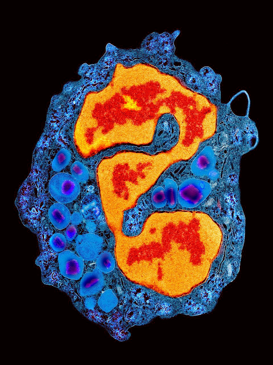
Our featured image, acquired by Rossana Melo, shows a human eosinophil isolated from the peripheral blood using a negative immunomagnetic selection method and prepared for transmission electron microscopy (TEM). The euchromatin and heterochromatin of the polylobed nucleus were pseudocoloured in red and orange respectively. Secretory granules, seen as large containers in the eosinophil cytoplasm, were highlighted in blue with their typical crystalline core in purple. The image was acquired with a Tecnai G2 Spirit Bio Twin ThermoFisher Scientific (former FEI) electron microscope.
Find out more about Rossana’s research below:
Research career so far: I’m a Professor of Cell and Molecular Biology at the Institute of Biological Sciences, Federal University of Juiz de Fora (UFJF), Brazil. I received my PhD and MS in Cell Biology from the Federal University of Minas Gerais (Brazil), post-doctoral training from Harvard Medical School, and held several visiting scientist positions at Harvard University (Beth Israel Deaconess Medical Center and Harvard T.H. Chan School of Public Health).
Current research: My research studies primarily aim to understand how cells from the immune system, especially eosinophils, respond to inflammatory situations and infectious diseases by investigating basic cellular mechanisms. My lab explores mechanisms of vesicular transport, cell secretion, endocytosis, cell death, and cell-cell and cell-microenvironment interactions.
Favourite imaging technique/microscope: I am particularly drawn to TEM. Application of conventional TEM in combination with quantitative assessments (quantitative TEM), molecular imaging (immunoEM), and three-dimensional (3D) electron tomography has consistently provided incredible insights into the intricate architecture and functionalities of immune cells, in the context of both health and disease.
What are you most excited about in microscopy? As a specialist in cellular ultrastructure, I’m continuously excited about the fascinating microscopic cell world. I’m delighted to mentor young scientists on their journey into this captivating world. I am also thrilled with the striking visual of cell images as a form of art. I have been using cell images, especially electron micrographs collected from many years of scientific research, to educate in the cell biology field and disseminate science through Science-Art exhibitions.
 (6 votes, average: 1.00 out of 1)
This approach is crucial as it provides detailed insights into the structural and functional aspects of immune cells. By utilizing advanced microscopy techniques, Rossana can unravel the complexities of cellular interactions in health and disease, paving the way for new discoveries and advancements in biomedical research.
Amazing image!
Very impressive!
Outstanding scientific art work .
Rosanna is a Co-PI on a project, the Bioimaging Network for the Advancement of Biomedical Research in Minas Gerais (BioIMG Net), which is funded by the Chan Zuckerberg Initiative (CZI) Expanding Global Access to Bioimaging Program. She and other members of BioIMG Net are introducing young students in underdeveloped regions of our state to the amazing beauty of science using images like this, as well as giving them the chance to manipulate microscopes at hands-on science fairs. Hundreds of students have participated in these outreach programs and always give the events great reviews.