Featured image with Jovan Brockett
Posted by FocalPlane, on 19 July 2024
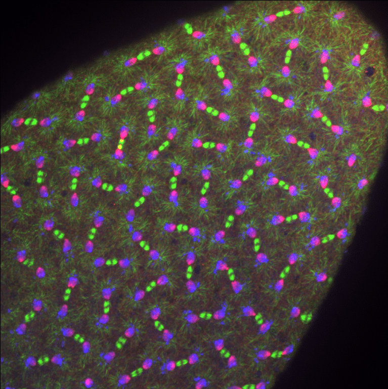
Our featured image, acquired by Jovan Brockett, shows a Drosophila melanogaster embryo undergoing syncytial nuclear divisions during early embryonic development. It was prepared using PFA fixation and hand dissected to remove the outer vitelline membrane. The image was captured using a Nikon Ti-E system fitted with a Yokagawa CSU-X1 spinning disk head, Hamamatsu Orca Flash 4.0 v2 digital CMOS camera, Perfect Focus system, Nikon LU-N4 solid state laser launch (15 mW 405, 488, and 561 nm) using high NA Apo TIRF oil immersion; ×40, 1.3 NA Plan Fluor oil immersion powered through Nikon Elements AR software on a 64-bit HP Z440 workstation.
Find out more about Jovan’s research below:
Research career so far: I have been in a research lab for seven years now. I spent three years as an undergraduate honor’s researcher at Oglethorpe University and four years as a Lead Research Specialist in the Lerit lab at Emory University School of Medicine.
Current research: As a Lead Research Specialist, I have my hand in a lot of different projects, which makes things even more exciting. My main research focus is on the early developmental period of Drosophila melanogaster embryos during which germline cells are the first to differentiate from the somatic body. Here, I focus on the centrosomes and microtubules to understand how this process happens and how defects in these processes can lead to developmental disorders. My secondary research focuses on how centrosomes affect melanoma metastasis and how a clinically identified SNP could contribute to renal cell carcinoma.
Favourite imaging technique/microscope: My favorite imaging technique is fixed-cell imaging because it allows me to be creative with the fluorescent probes and design how I want the structures to look. I would like to learn how to do expansion microscopy or super-resolution microscopy so I can look at these objects even closer with more vivid colors.
What are you most excited about in microscopy? I am most exciting about the future of Quantum Enhanced nonlinear Microscopy, which utilizes quantum properties, such as entanglement, to improve the resolution and sensitivity of nonlinear microscopy. This technique improves resolution, sensitivity, signal: noise, and reduces photobleaching. Additionally, you do not have to use fluorescent probes and can rely on the natural refraction of polarized light from the object in some cases.
 (2 votes, average: 1.00 out of 1)