Kidney elastic fiber network changes in Marfan’s Syndrome
Posted by Mario Elias Ortega Sandoval, on 19 July 2024
by de Souza RB*, Meek KM#, Lewis PN#, Pereira LV*.
*Department of Genetics and Evolutionary Biology, University of São Paulo, Brazil
#StructuralbBiophysics Research Group, School of Optometry and Vision Sciences of the United Kingdom, Cardiff University, UK
Challenge
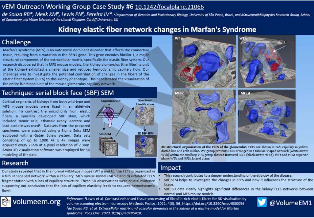
Marfan’s syndrome (MFS) is an autosomal dominant disorder that affects the connective tissue, resulting from a mutation in the FBN1 gene. This gene encodes fibrillin-1, a major structural component of the extracellular matrix, specifically the elastic fiber system. Our research discovered that in MFS mouse models, the kidney glomerulus (the filtering unit of the kidney) exhibited a smaller size and reduced hemodynamic capillary flow. Our challenge was to investigate the potential contribution of changes in the fibers of the elastic fiber system (FEFS) to this kidney phenotype. This necessitated the visualization of the entire functional unit of the mouse glomerulus capillary network.
Technique: serial block face (SBF) SEM
Cortical segments of kidneys from both wild-type and MFS mouse models were fixed in an aldehyde solution. To contrast the microfibrils from elastic fibers, a specially developed SBF stain, which included tannic acid, ethanoic uranyl acetate and lead acetatewas used1. Datasets from the prepared specimens were acquired using a Sigma Zeiss SEM equipped with a Gatan 3view system. Data sets consisting of up to 1000 4k x 4k images were acquired every 75nm at a pixel resolution of 7.3nm. Amira 3D visualization software was employed for 3D modeling of the data.

Research
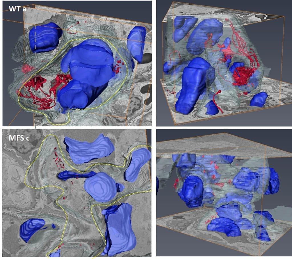
Our study revealed that in the normal wild-type mouse (WT a and b), the FEFS is organized in a tubular-shaped network within a capillary. MFS mouse model (MFS c and d) exhibited FEFS fragmentation with a loss of capillary structure. These 3D observations were crucial evidence supporting our conclusion that the loss of capillary elasticity leads to reduced hemodynamic flow2.

Impact
• This research contributes to a deeper understanding of the etiology of the disease.
• SBF-SEM helps to investigate the changes in FEFS and how it influences the structure of the tissue.
• SBF 3D data clearly highlights significant differences in the kidney FEFS networks between normal and MFS mouse models.
References
1Lewis et al. Contrast-enhansed tissue processing of fibrallin-rich elastic fibres for 3D visulisation by volume scanning electron microscopy Methods Protoc. 2021, 4(3), 56; https://doi.org/10.3390/mps4030056
2de Souza RB, et al. Extracellular matrix and vascular dynamics in the kidney of a murine model for Marfan syndrome. PLoS One. 2023. 9;18(5):e0285418.
Poster


 (1 votes, average: 1.00 out of 1)
