Featured image with David Gaboriau
Posted by FocalPlane, on 11 October 2024
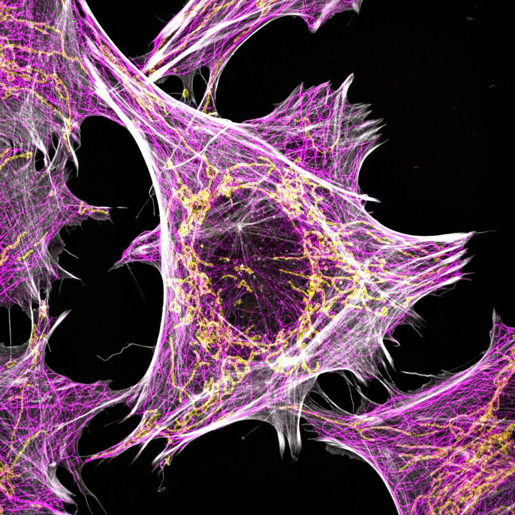
Our featured image, acquired by David Gaboriau, shows human fibroblasts with mitochondria (Tom20) in yellow, alpha tubulin in pink and actin in grey. It’s a 3D-SIM image taken on a Zeiss Elyra PS.1, reconstructed in Zen and presented as a maximum intensity projection.

Find out more about David’s research below:
Research career so far: I’m originally a biochemist and cell biologist and have used various imaging techniques and bioimage analysis for my research. I did my PhD at the Babraham Institute in Cambridge, working on protein-protein interactions during fertilisation. After completing my PhD, I worked in drug discovery at KuDOS Pharmaceuticals/AstraZeneca on a PARP inhibitor called Olaparib, which is now used to treat breast, ovarian, pancreas and prostate cancers in patients with faulty BRCA genes. I then joined Prof Laura Itzhaki’s group in Cambridge to work on DNA repair and protein folding, looking at the BRCT domains of BRCA1. Next, I went to Galway in Ireland for two post docs at the Centre for Chromosome Biology, with Prof Ciaran Morrison on centrosome biology and DNA repair, and with Prof Corrado Santocanale on Claspin, CDC7 and the regulation of the cell cycle. In 2015, I moved to London to join the Facility for Imaging by Light Microscopy (FILM) at Imperial College as a Microscopy Specialist.
Current research: My current post as Deputy Manager and Microscopy Specialist at FILM involves looking after the facility in our South Kensington campus and providing expert training and assistance in light microscopy to our users, who come from all Faculties and Departments at Imperial College. We start with a chat about the project and the experiments, then we train and accompany the user to help them get the best quality images for their research. I also train users on the basics in bioimage analysis. My favourite part of the job is training and teaching, and seeing the user get confident on the microscope, and in return we get to learn new science about their projects. If possible, I love to collaborate with users, either on advanced imaging or bioimage analysis. It’s important for core facility staff to collaborate, for personal and scientific development, and to keep up with these two fast evolving research fields!
Favourite imaging technique/microscope: There are a few, and each is most suitable to address a specific question, so it depends. In the facility, we have ‘workhorse’ systems that a lot of users will use, Zeiss Axio Observer widefields and Leica SP8/Stellaris confocals, and they are excellent. For the majority of projects, these standard systems work very well. For super-resolution, I love the Zeiss Elyra PS.1 and NIS Elements and JOBS for Nikon systems. For high-content imaging, I had an Operetta in the lab in Galway that I liked a lot.
Regarding bioimage analysis, I love FIJI and Icy, and as the field moves to using Python, the entry level gets higher for biologists without coding skills. Most users ask for easy-to-use solutions, and bioimage analysis can be quite complicated, so our role is to educate, advise, and point users towards resources that will help them. A great place to start is the image.sc forum! These times of high technological innovation are very interesting to witness and participate in.
What are you most excited about in microscopy? In the last 10 years especially, bioimage analysis has really developed, and it seems to go faster and faster. For this, NEUBIAS and others have been amazing in fostering a generation of bioimage analysts and putting this new career on the science map. The rise of AI in particular is very impressive, new techniques and applications come out nearly every day. It’s important to remember that not all projects and users need it, but for some applications it’s quite fantastic.


 (1 votes, average: 1.00 out of 1)