Vote for your favourite image
Posted by FocalPlane, on 11 March 2025
We are delighted to unveil the shortlisted images in the Node–FocalPlane image competition. Thanks to everyone that entered, we received so many fantastic images, and congratulations to all the finalists. You can vote for your favourite image using the poll at the bottom of this page.
The shortlisted images will also be displayed in our gallery at the Biologists @ 100 conference, which is being held in Liverpool from 24-27 March. We’ll add up the votes from FocalPlane, the Node and our conference delegates, and announce the winners at the end of the conference.
Voting will close at 23:59 GMT on Wednesday 26 March.
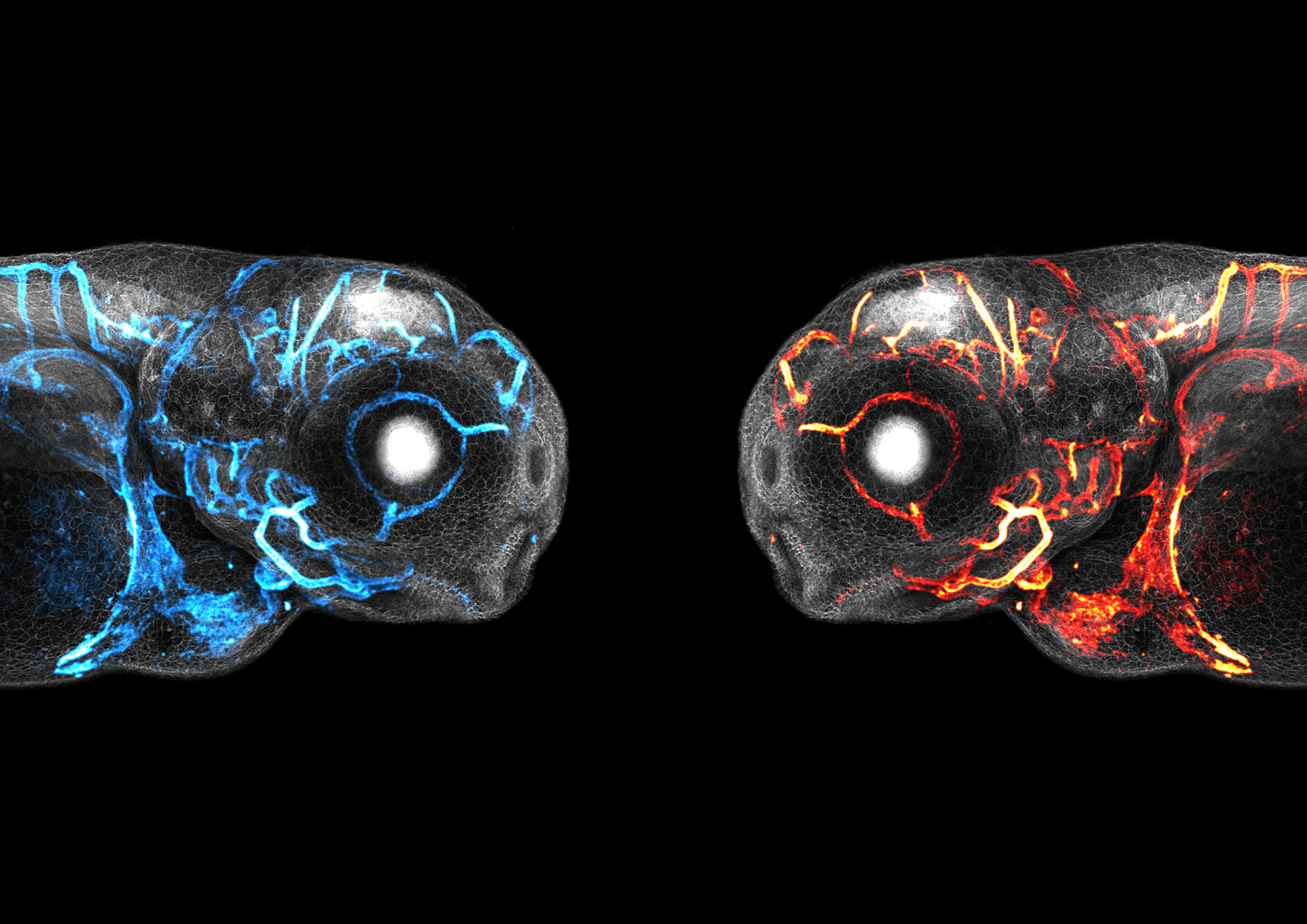
1. A song of ice and fire – Aaron Scott
The plasma membrane of every cell in these 2-day old larval zebrafish is fluorescently labelled and shown in grey. The endothelial cells and the blood vessels they form are shown in cyan or red. Imaged on a Leica SP8 AOBS confocal laser scanning microscope and reconstructed using ImageJ.
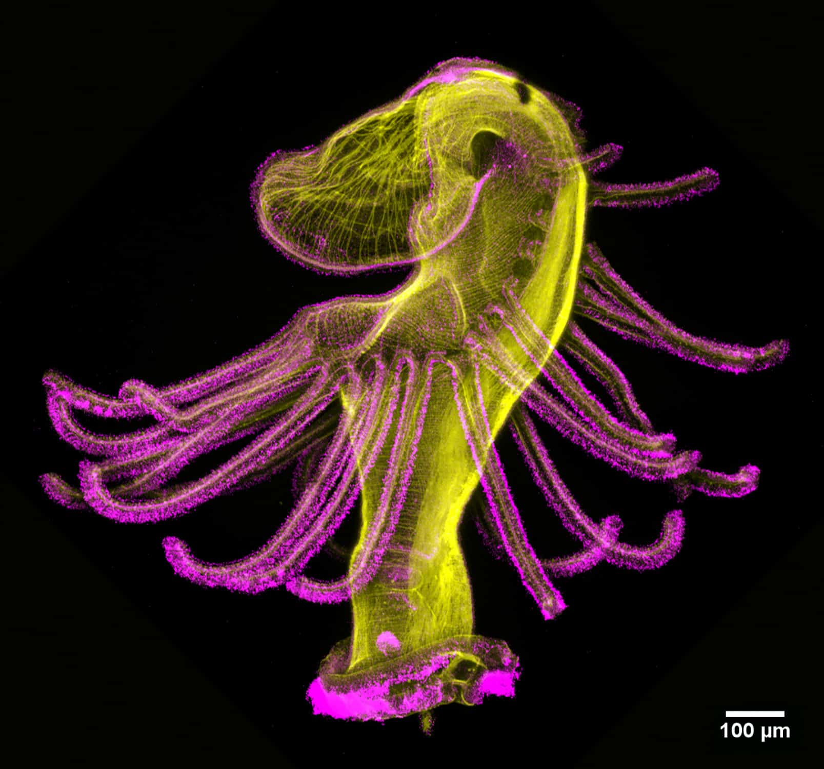
2. Dancing actinotroch – Allan Carrillo-Baltodano
Actinotroch larva of a phoronid worm with phalloidin shown in yellow and acetylated tubulin in magenta. Imaged with a Zeiss LSM 800 at 10 x magnification.
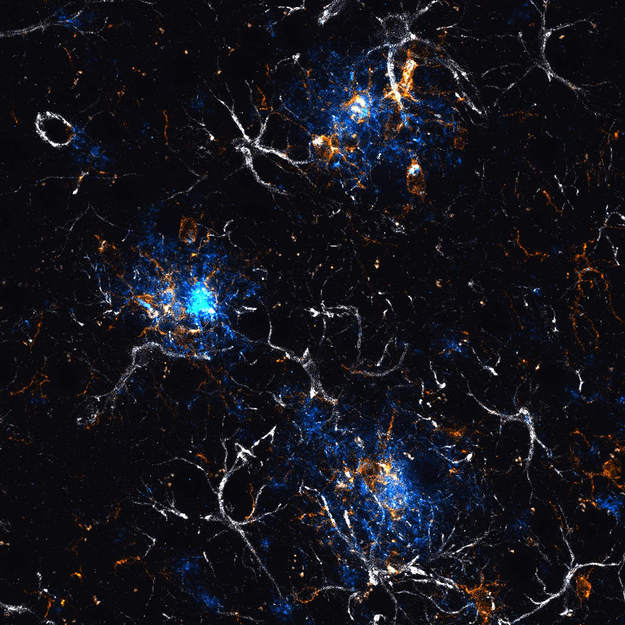
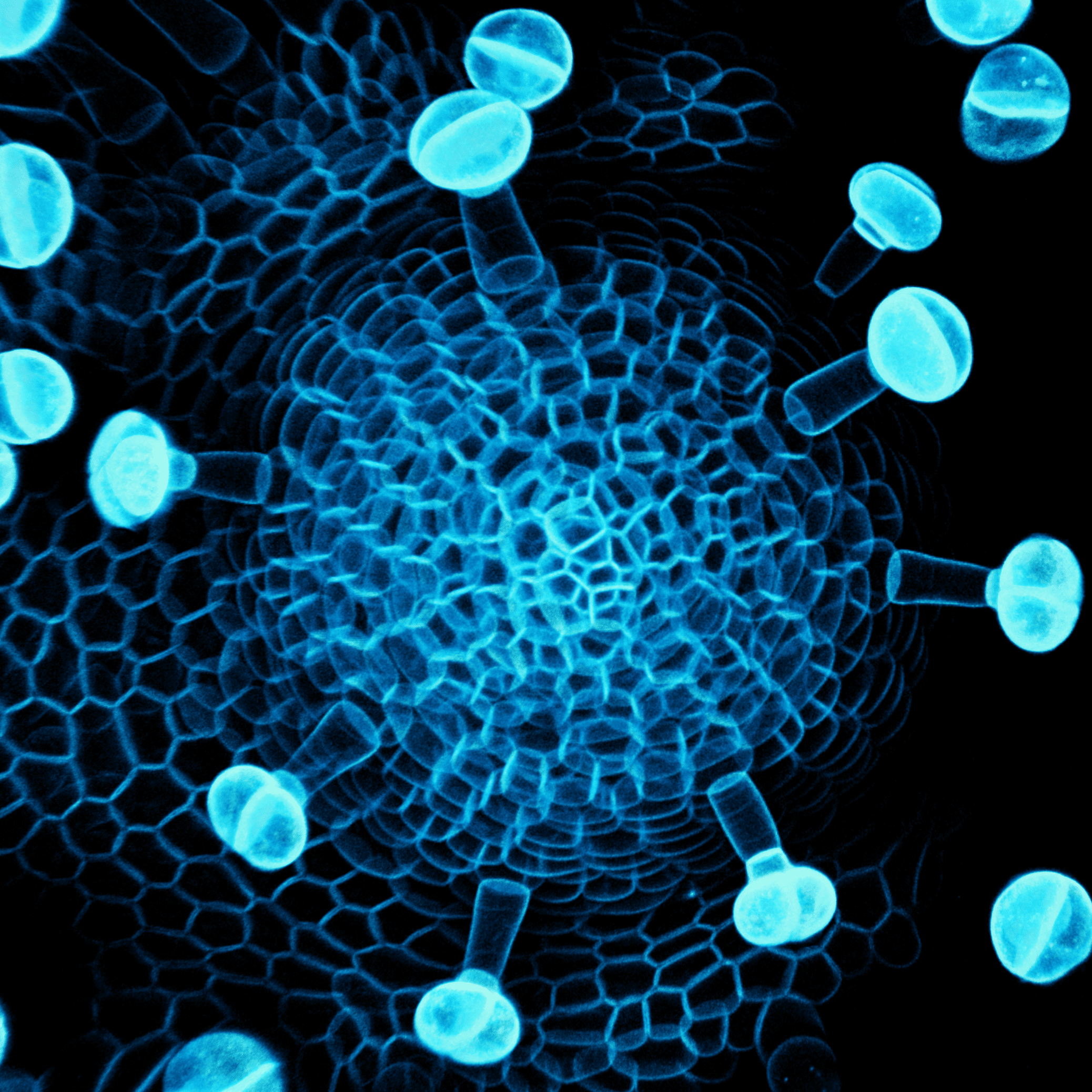
3. The glial response to amyloid skies – Andrew Octavian Sasmita
Confocal maximum projection image of several amyloid-β plaques (blue, 6E10) surrounded by microglia (gold, Iba1) and astrocytes (white, GFAP) in the cerebral cortex of a 6-month-old female APPNLGF mouse model of amyloidosis. Imaging was done with a Zeiss LSM 800 Airyscan confocal microscope and processed with the Zen imaging software.
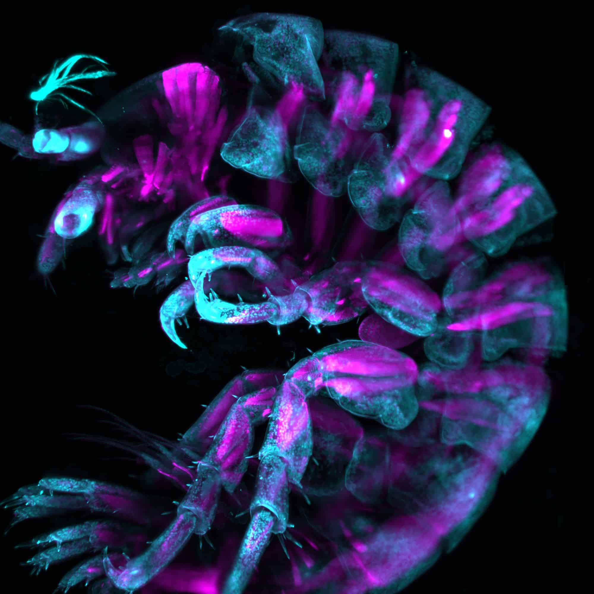
4. A mystery amphipod – Camila Weiss, José Palma and Marina Cuenca
Lateral view of an unknown species of chilean amphipod labelled with DAPI (cyan) and phalloidin (magenta). Imaged using a Zeiss Lightsheet 7 at the Quintay developmental biology course in 2023 and processed with Fiji.
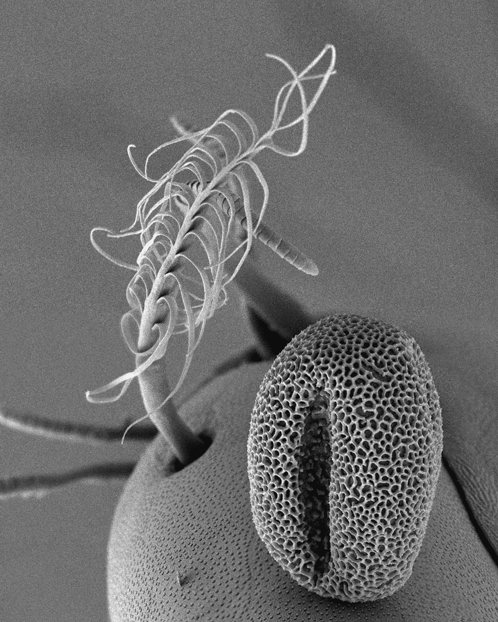
5. A beautiful contamination – Çağrı Çevrim
A scanning electron micrograph (SEM) of a Parhyale hawaiensis limb, showing an external mechanosensory organ – a plumose seta – in the background. In the foreground, a pollen grain, possibly from a Platanus tree, has contaminated the sample.
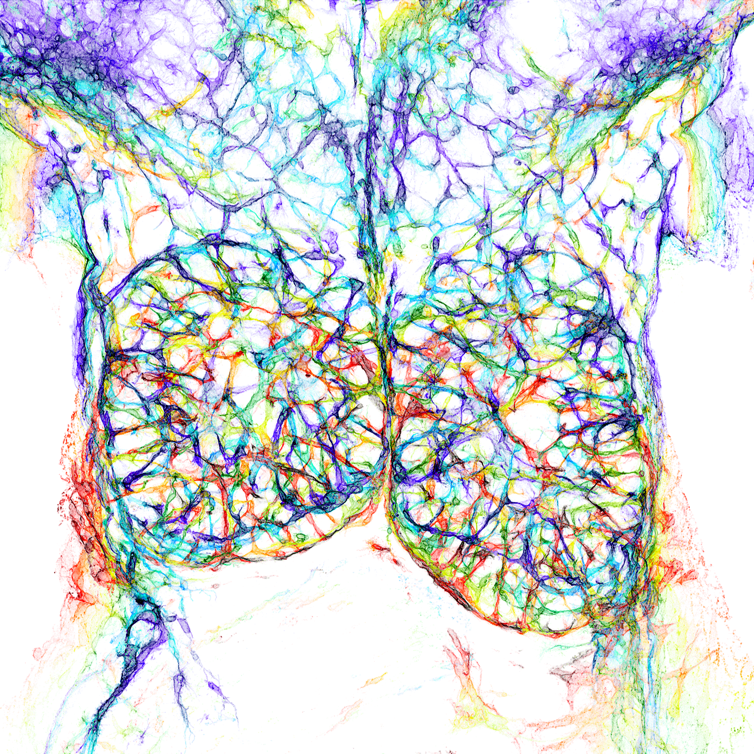
6. Thymus in the spotlight – David Grainger
A colour-coded depth projection of the blood endothelial cells of the E14.5 mouse embryonic thymus and surrounding structures. A 200 μm-thick vibratome section was immunostained for endomucin and imaged on a Zeiss LSM980 confocal microscope and depth-encoded using a rainbow LUT before performing a maximum intensity projection.
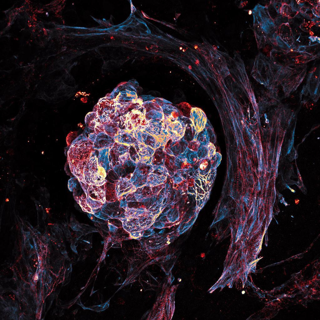
7. Beat - Ioakeim (Makis) Ampartzidis
Mature beating cardiomyocyte cell cluster from human induced pluripotent stem cells. Cells were grown for a total of 20 days and stained positive for cardiac Troponin (Hot Blue) and actinin (Hot Red) markers. The image was acquired at the Veneto Institute of Molecular Medicine (VIMM), in Nicola Elvassore’s lab, using an upright LSM900 ZEISS microscope and LUTs adjusted using Fiji software.
8. Who’s active? – Julia Peloggia de Castro
The image depicts a zebrafish embryo at 9 hours post-fertilisation on a lateral view. Cells are stained with MitoTracker, which labels active mitochondria, and cell membranes are labelled in cyan with a EGFP transgenic membrane tag. Image was taken using a 20x objective on a spinning disk confocal microscope.
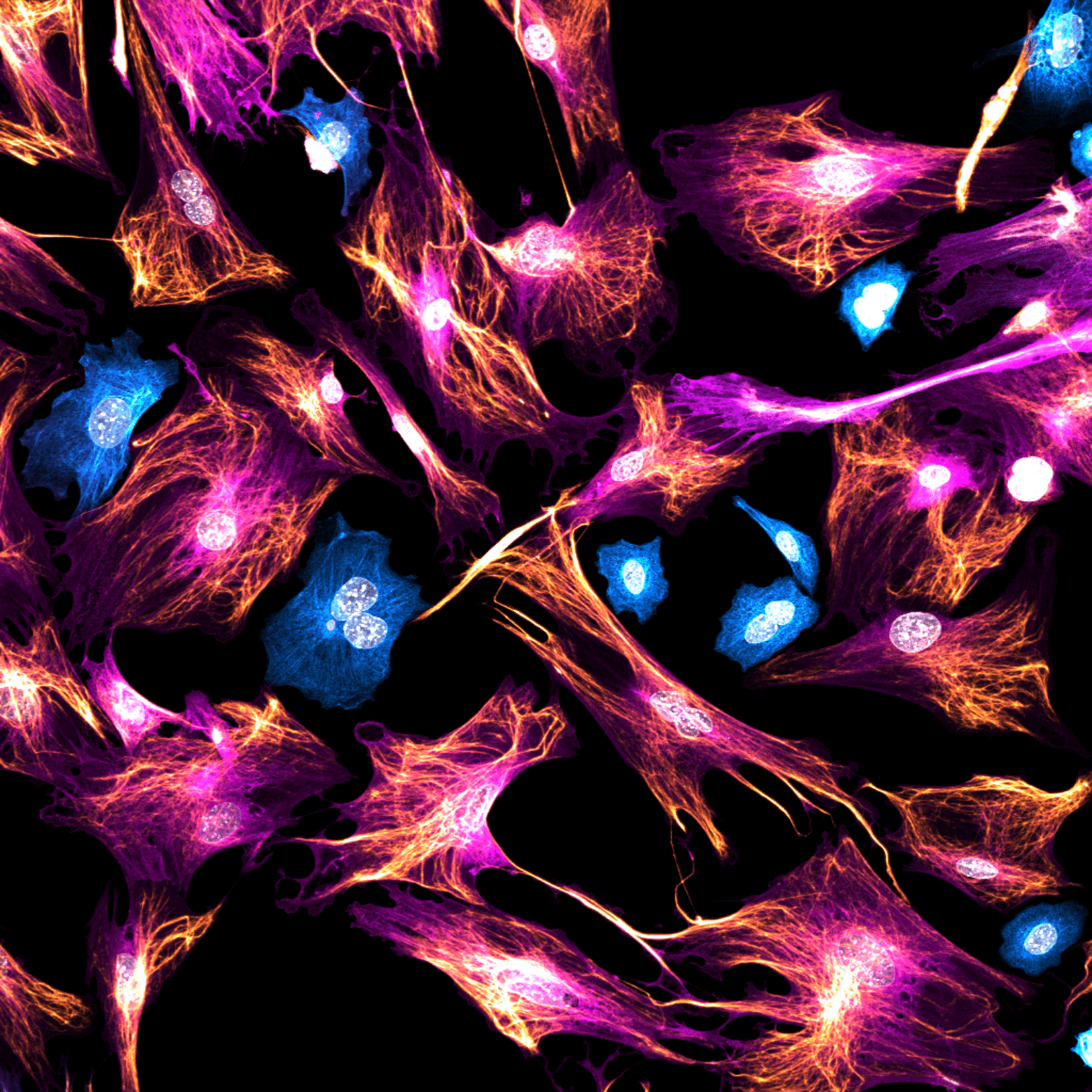
9. Unexpected guests – Krystyna Gieniec
2D culture of mouse mammary fibroblasts stained for Acta2 (magenta) and Vimentin (gold), with some contaminating epithelial cells stained for pan-Cytokeratin (cyan). Image acquired using a Leica Stellaris 8 confocal microscope.
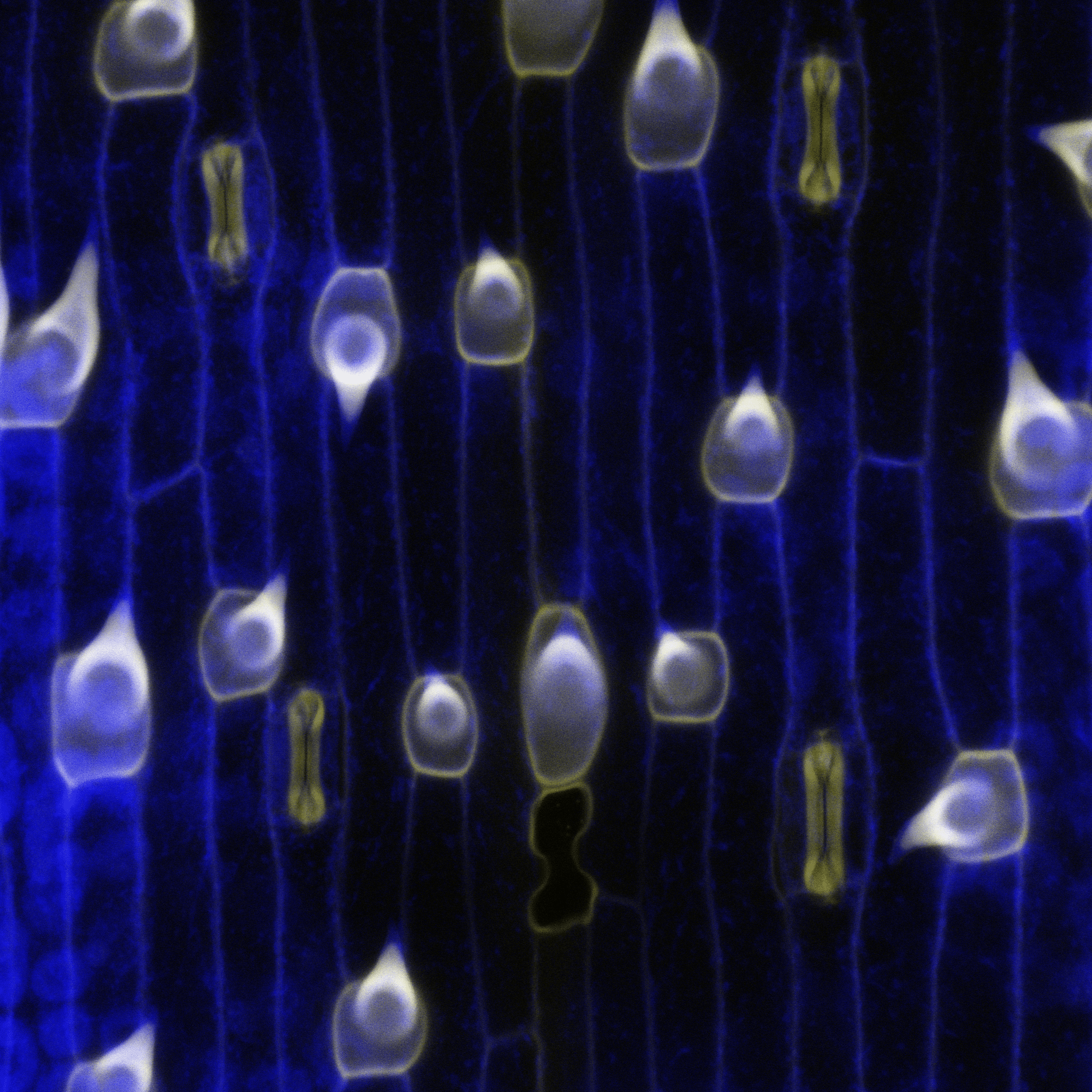
10. The plant-atmosphere interface that feeds the world – Lea Berg and Michael Raissig
Mature epidermal cell types in a grass leaf of the emerging developmental model system Brachypodium distachyon. Cell outlines are blue, which is plant cell wall UV-autofluorescence. In yellow is stained lignin, a secondary cell wall modification that can be found in the hair cells (‘shark-tooth’-shaped) and the stomatal guard cells (‘dumbbell’-shaped). Imaged by confocal microscopy and processed in Fiji.
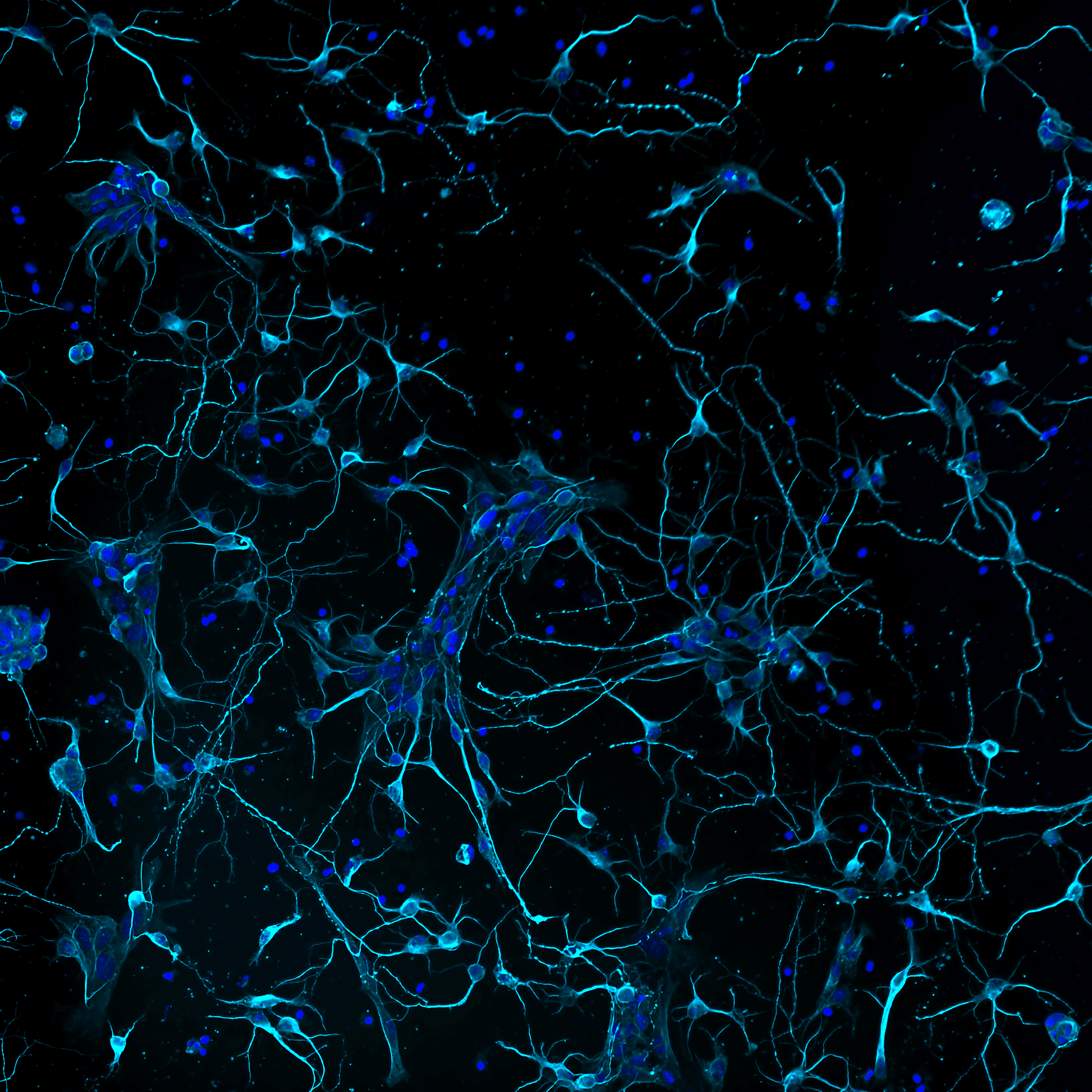
11. Plenty of fish in the sea – Ludovica Altieri
Murine primary cortical neurons developing interconnections, stained with neuronal tubulin (cyan) and DAPI (blue). Imaged on a Nikon microscope implemented with a CrestOptics confocal spinning disk module with post-processing using NIS Elements AR by Nikon. Acquired at the IBPM Institute of Molecular Biology and Pathology – CNR National Research Council of Italy, c/o Department of Biology and Biotechnology “Charles Darwin” – Sapienza University of Rome.
12. Invisible architects – Maik Bischoff
Drosophila hydei testis musculature stained with phalloidin to label F-actin (blue), anti N-Cadherin (orange/gold) to mark cell-cell junctions between muscle cells and DAPI to stain nuclei (purple). Autofluorescence (orange/gold) makes the trachea visible. Imaged with a Zeiss 980 confocal microscope with Airyscan 2. The image was processed in Zen Blue, and LUTs (by KTZ) applied in Fiji with further modifications in Photoshop.

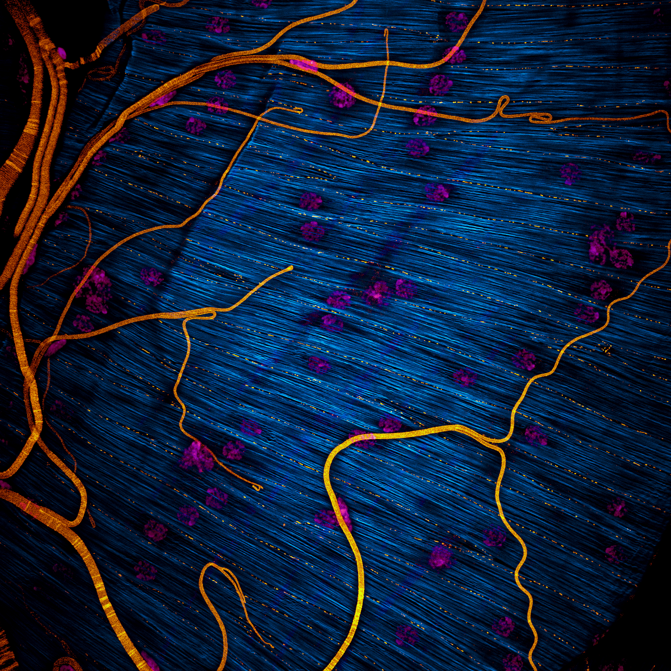
13. Breath of the water – Mathieu Preußner
Lateral view of the overlying gill arches in 1-month-old Danio rerio expressing endothelial kdrl:mCherry. Clarity-based tissue clearing of the sample enabled comprehensive image acquisition using a Nikon Ti spinning disk system. In ImageJ, the hyperstack was modified using a temporally colour-coded lookup table.
14. Pin shoot – Min Ya
Maximum projection of confocal stacks of a mutant Mimulus parishii shoot apex with cells labelled with a plasma membrane marker. The shoot apices of this plant can grow but are unable to produce any organs, resulting in a phenotype that resembles a pin.
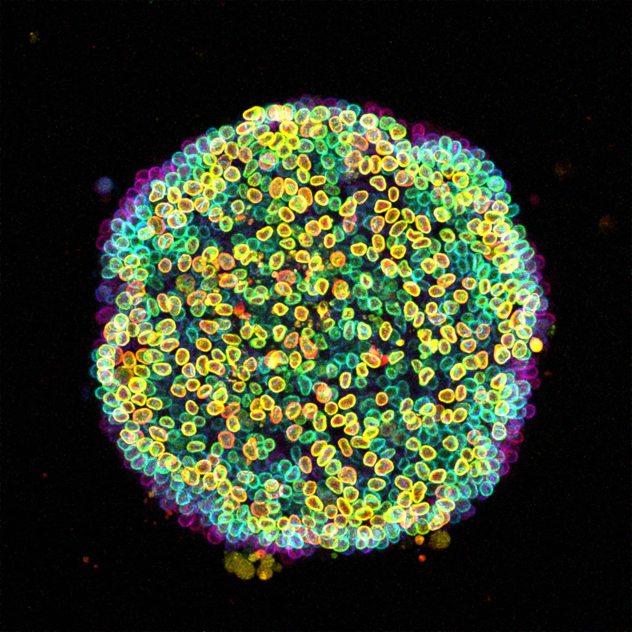
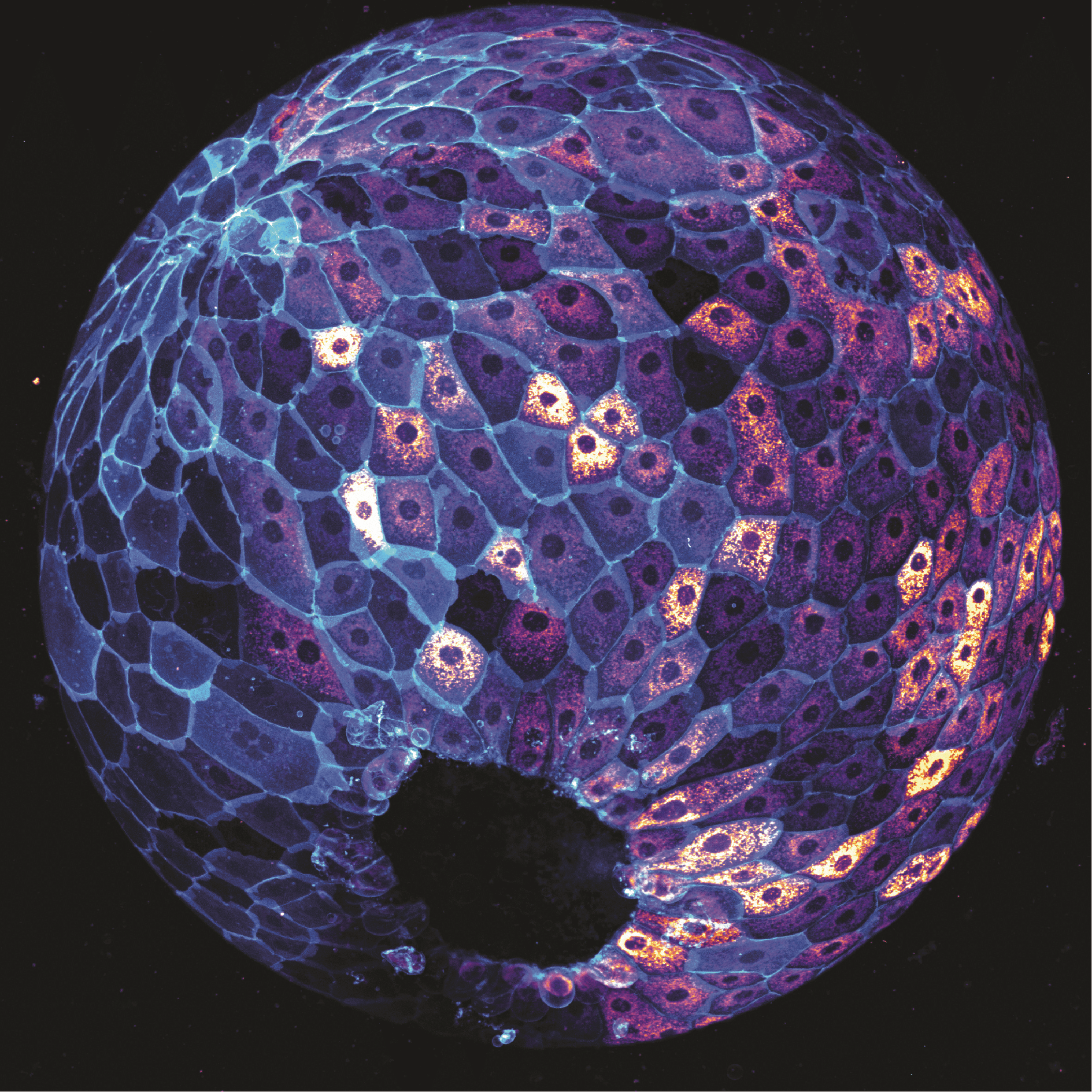
15. Cell-estial bloom – Özge Özgüç
A ‘Cell-estial bloom’ of human induced pluripotent stem cells (hiPSCs) flourishes on a micropatterned island. This image presents a colony of live hiPSCs, with fluorescently labelled Lamin B delineating the nuclear lamina within each cell. Acquired with a Zeiss LSM 880 Airyscan microscope, this maximum intensity projection is enhanced with depth-coded colouring to reveal the captivating three-dimensional landscape.
Thank you for voting
 (44 votes, average: 1.00 out of 1)
Great images! Science and art in a nutshell
Great image with good contrast.
All these images are magnificent ! The choice is impossible, so I followed my impulsion.
Good pic
Amazing images, congrats to everyone!
As an amyloid researcher I am always fascinated by the glial response and its beauty
Great neurons!
Fantastic images, thank you everyone for sharing the beauty of your work!
Fsntastics
amazed by the science creating art !
Mesmarizing shots, I am thrilled to see intense beauty in science images!
Good luck to all participants!
Buda bir sanat ve çok güzel
Buda bir sanat ve çok güzel bence
Incredible and magnificent works,arts of science..I wish success to all participants, and appreciate their hard work.
The beauty of the science is simply stunning!
The Node–FocalPlane competition celebrates the beauty of scientific imaging, and Özge Özgüç’s ‘Cell-estial Bloom’ perfectly embodies this with its stunning depiction of hiPSC organization.
This competition highlights the power of microscopy in uncovering cellular landscapes, and Özge’s work stands out as a striking fusion of scientific precision and artistic brilliance.
Özge Özgüç’s image captures the essence of the Node–FocalPlane competition—transforming complex biological structures into breathtaking visuals that inspire both scientists and the public.
Marvellous