Featured image with Subhajit Karmakar
Posted by FocalPlane, on 11 April 2025
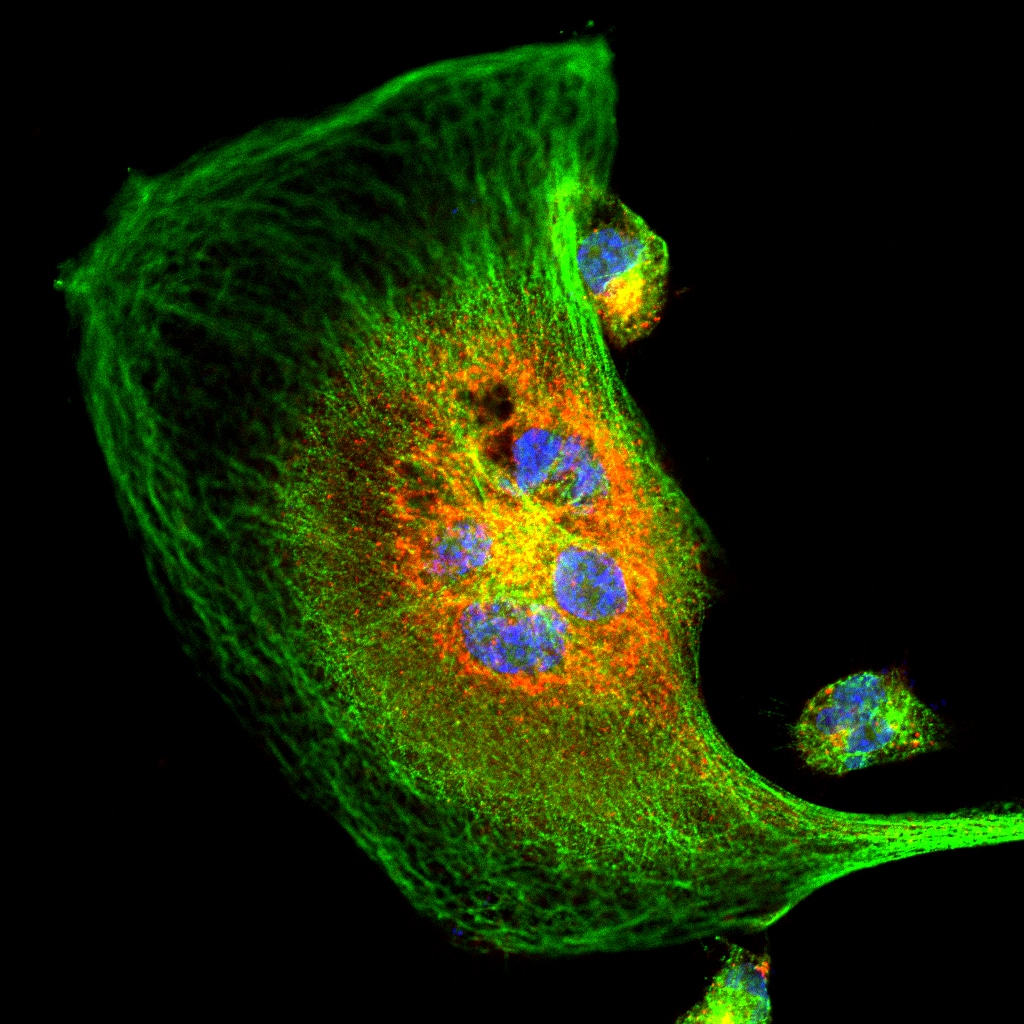
Our featured image, acquired by Subhajit Karmakar, depicts a dividing MDA-MB-231 cancer cell that has been stained to visualise the localisation of two key proteins: CK2, a highly oncogenic kinase, and an E3 ligase, a tumour suppressor. The study focusses on the variation in CK2 localisation under serum-starved conditions, aiming to understand its cellular distribution in response to nutrient deprivation. To prepare the sample, the cells were fixed with 4% paraformaldehyde to preserve their structure. They were then incubated with primary antibodies specific to CK2 and the E3 ligase, followed by secondary antibodies conjugated to Alexa Fluor dyes for fluorescence detection. Finally, the cells were mounted with DAPI to stain the nuclei, allowing for clear visualisation of the cell’s overall structure and the specific localisation of the proteins. This approach provides valuable insights into the dynamic behaviour of these proteins in response to cellular stress.
Discover more about Subhajit’s research
Research career so far: My research career so far has been a fulfilling journey of exploring the intricate mechanisms underlying cancer biology. Currently, I am a 5th-year PhD student at CSIR-Indian Institute of Chemical Biology (IICB), working under the guidance of Dr. Mrinal K. Ghosh. My academic journey began with a Master of Technology (M.Tech) degree from IIT Kharagpur, where I developed a strong foundation in molecular and cellular biology. Driven by a passion for understanding cancer progression, I joined IICB to delve deeper into the signalling transduction mechanisms that govern key molecular pathways in cancer.
Current research: At IICB, my research focusses on unravelling the complex molecular mechanisms that regulate cell proliferation and inhibit apoptosis, two critical processes in cancer development. Specifically, I investigate how dysregulation in these pathways contributes to tumour growth and survival. My work combines advanced molecular biology techniques, cell-based assays, and imaging methodologies to dissect the signalling networks involved in cancer progression. Through this research, I aim to contribute to the identification of potential therapeutic targets that could pave the way for more effective cancer treatments in the near future.
Favourite imaging technique/microscope: My favourite microscope is the ZEISS LSM 990.
What are you most excited about in microscopy? I am most excited about the ability of microscopy to reveal the dynamic architecture of cellular proteins and how they shape cell structure and function. Specifically, I find it fascinating to visualise and analyse the internal localisation of proteins within cells and understand how their spatial organisation regulates complex signalling networks. In the context of cancer, microscopy allows us to observe how protein dynamics and localisation contribute to mechanisms like uncontrolled cell proliferation, metastasis and inhibition of apoptosis. By capturing these intricate details, we can uncover critical insights into the molecular underpinnings of cancer progression and identify potential therapeutic targets.
 (4 votes, average: 1.00 out of 1)