An Interview with Dr. Rashmi Priya
Posted by Subhajit Dutta, on 1 July 2025
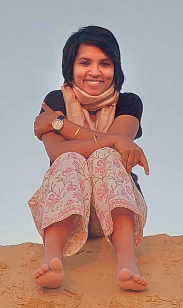
Welcome to the FocalPlane interview series, where we spotlight groundbreaking Asian-origin scientists pushing the boundaries of cell biology and microscopy. Today, we are delighted to feature Dr. Rashmi Priya, a group leader at the Francis Crick Institute (UK). In this insightful interview, Dr. Priya shares her unconventional journey into science, her fascination with organ morphogenesis, and her dedication to fostering interdisciplinary collaboration. Join us as we delve into the exciting world of developmental biology and microscopy through the lens of Dr. Priya’s expertise. Dr. Rashmi Priya leads a research group at the Francis Crick Institute, focusing on understanding the fundamental principles governing organ development. With a background in cell biology and expertise in live imaging, her lab investigates how organ form and function emerge robustly during morphogenesis. Dr. Priya employs an interdisciplinary approach, combining genetics, biophysics, and advanced microscopy to unravel the intricate mechanisms that orchestrate tissue formation.
Q: How did you start your career in science? What sparked your interest in cell biology and microscopy?
A: My path into science wasn’t exactly planned—it was more of a happy accident. Having grown up in India in a modest socioeconomic setting, science as a career wasn’t really on the table. While research as a career is becoming more democratic, it still often requires a certain level of privilege and access.
For me, it was a news article about the Human Genome Project—and a comment by thePresident of India, Dr. APJ Abdul Kalam—that really caught my attention and got me wondering: What is research? Who are the people doing it? And how does one become a scientist?
Things really shifted for me during a summer research fellowship at the Saha Institute of Nuclear Physics. It was fully funded by the Department of Science and Technology, and it gave me my first real taste of lab life. That experience opened my eyes to what research actually involves—and that it could be a viable career path, with many possibilities. I didn’t set out to study cell biology or microscopy. During my master’s internship at Jawaharlal Nehru University, I worked on the malaria parasite, which deepened my interest in biology. Then, during my time at Tata Memorial Hospital, I looked at how cancer cells stick to each other and spread, which got me thinking more about cellular behaviour. But it was a personal move to Australia that really pivoted things for me. I got an opportunity to do PhD in Alpha Yap’s lab, and that’s where I got into cell biology and microscopy—studying how epithelial cells hold together and maintain their structure. That’s when it really clicked for me; I wanted to understand how thousands of individual cells come together to build intricate functional organs and then a fully grown organism.
Q: What were some of your key takeaways from your PhD experience in Australia?
A: My time in Alpha Yap’s lab was truly transformative. It wasn’t just about doing experiments—it was about how to ask creative questions, design thoughtful experiments, and troubleshoot when things didn’t go as planned. Alpha was a fantastic mentor who created a supportive and intellectually rich environment. The lab’s international mix also exposed me to different ways of thinking and doing science. It was also my first experience in a world-class research institute—with impressive infrastructure, a well-structured PhD program, and so many training opportunities. I honestly felt a bit spoiled and incredibly privileged to be in that environment. I think that awareness always stayed with me—I knew I had to make the
most of the opportunity.
Q: What drew you specifically to the world of organ morphogenesis, especially using the
heart as a model system?
A: By the end of my PhD, I knew I wanted to explore the complexities of how cells behave in living organisms—especially during organ development. Zebrafish felt like the ideal system for this. I was particularly drawn to the heart. It’s an engineering marvel! What starts as a simple linear tube transforms into an intricately structured pump that beats continuously to sustain life. How has nature designed such an efficient, robust system? And, how does something so complex come together during development?
Studying the heart brought its own set of challenges—zebrafish hearts beat about three times per second, so imaging such a fast-moving organ and capturing cellular behaviours at the right scale isn’t straightforward. But that’s exactly what made it exciting. It pushed me to think beyond the obvious. Also, surprisingly, despite its importance, heart morphogenesis is still relatively understudied, and there is still a lot to explore.
Q: You mentioned the importance of mentorship. How do you find mentors?
A: Finding the right supportive environment is absolutely crucial. I lean on my mentors a lot! I chat regularly with my peer mentors about all the ups and downs of academia — whether it’s publications, grant rejections, people management, or just needing to vent. I’m always bothering my senior colleagues for advice and, honestly, just for encouragement too, because we all need that. Just because you’ve become a PI doesn’t mean you don’t still need to hear, “You’re doing well.” Those words of encouragement really do matter.
Q: As a PI, how do you foster an environment in your lab that encourages interdisciplinary thinking and collaboration?
A: I really believe that answering complex biological questions needs an interdisciplinary approach. In our lab, we create an open, supportive space where everyone feels comfortable sharing ideas. We actively recruit people from diverse backgrounds, which brings a great mix of skills and perspectives. Our projects naturally span areas like imaging, genetic
engineering, and biophysics, so collaboration happens quite organically. Regular journal clubs on different topics and lab meetings also help us stay curious and engaged with science beyond our own fields.
Priya Lab: Organ Morphodynamics Laboratory
Q: Establishing a new lab comes with its own set of challenges. Could you share some of the hurdles you faced while setting up your lab, and how did you overcome them?
A: Starting a lab is both exciting and a bit overwhelming. As a new PI, you’re suddenly juggling things you were never really trained for — from budgets and equipment to mentoring and grant writing. Doing all that while settling into a new country as an international scientist adds an extra layer of challenge. One of the toughest parts was building a team from scratch. It took time and care to bring together people with the right mix of personalities and research interests. Thankfully, I’ve had amazing support from the Francis Crick Institute. They’ve built a great infrastructure for new PIs, which made those early days a lot more manageable.
Q: What makes working at the Crick Institute unique and how does it impact your research?
A: The ‘six plus six’” model, which offers 12 years of core funding, is such a fantastic initiative. It really gives group leaders like me the space to focus on building our research programs and chasing ambitious ideas, without the immediate pressure of securing external funding. That kind of freedom to explore bold, creative directions is absolutely invaluable, especially when you’re just getting a lab off the ground. Of course, grant writing is still very much part of the job, but having that core funding means we can take a more balanced approach. It lets us tackle those high-risk, high-reward questions while also keeping momentum on more established projects.
Q: What are some of the exciting projects that your team is currently working on?
A: There’s a lot going on when a single fertilized egg turns into a full-fledged organism. Cells are dividing, migrating, and taking on specific roles; tissues and organs are forming at just the right time and place; and molecular signals are everywhere. All of this somehow results in a structured body plan made up of highly complex organs with incredibly
specialised and robust functions. It’s absolutely mind-blowing when you think about it. In our lab, we’re trying to understand how functional organs are built during development. We use the zebrafish heart as our main model system to address this fundamental question—but the real goal is to uncover the general rules of morphogenesis that apply more
broadly across diverse tissues and organs. We recently wrote a review article highlighting these general rules.
Organ morphogenesis is a deeply multiscale problem. An organ is so much more than just a collection of cells. The shapes and structures that organs take on during development aren’t something you can simply predict from looking at genes or proteins alone. Patterns emerge across scales—cellular, tissue, mechanical—and that’s where things get really interesting.
We have several projects running that dive into different aspects of heart morphogenesis. One project looks at how the mechanical forces generated by the beating heart, this constant stretch and compression, affect the nuclei of heart muscle cells. These cardiomyocytes endure mechanical stress every second of their existence, yet their nuclei stay intact and protect the genome. We’re working to uncover how that happens, which could offer insights into aging and heart disease.
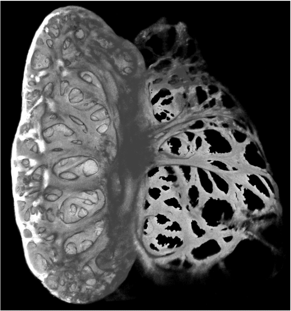
A mature zebrafish heart, 20 days post fertilization, showing the intricate meshwork architecture of ventricle and atrium.
Credit: Marie-Christine Ramel, Senior Laboratory Research Scientist, Priya Lab
Scaling up, we’re also looking at how a simple, epithelial-like primitive heart tube gives rise to the intricate 3D meshwork called trabeculae that helps the heart pump efficiently.
And something we recently discovered has been especially exciting: it turns out that the heart actually uses fractures or defects in the extracellular matrix to help pattern the myocardial wall. It’s fascinating—when you think of morphogenesis, you often think of perfection and precision, and yet here we see the system using what looks like failure or damage as a tool. That kind of unexpected insight is what keeps us motivated.
A beating zebrafish heart, 5 days post fertilization, myocardial cell membrane labelled in grey, red blood cells labelled in red.
Credit: Toby Andrews, postdoc, Priya lab
Q: What is your philosophy when it comes to nurturing the next generation of researchers?
A: I really believe in individualized mentorship — everyone comes with their own strengths, challenges, and goals. My job is to adapt to what each person needs, create an environment where they feel supported to explore their ideas, and help them leave the lab as confident, capable scientists. I keep communication open and offer feedback that’s timely and honest. For me, mentorship is a long-term investment. It’s not just about guiding them in the lab, but also helping them believe in their potential and prepare for future opportunities. I’m still learning how to balance the yin and yang of it all, but it’s one of the most rewarding parts of the job.
Q: How do you think the field can become more inclusive and supportive?
A: We’ve made substantial progress on diversity and inclusion, but it’s not enough to simply bring underrepresented groups to the table — we need to make sure they feel valued, supported, and able to thrive. That means challenging the systems and structures that continue to create barriers, and most importantly, celebrating the wide range of leadership
styles that people bring.
One area that often gets overlooked is access to informal networks — those behind-the- scenes connections built through social interactions that can be critical for career growth. We need to create more spaces for underrepresented groups to access those networks and the support that comes with them.
There is considerable emphasis on training women and academics from underrepresented groups on how be assertive and advocate for themselves. But we also need to change the system which puts these unnecessary barriers on them and expects them to fit into a certain mould. This will be a long road, but each step taken will contribute towards creating a truly inclusive scientific community.
Q: As a successful Asian woman in science, what advice would you give to young scientists who aspire to make a mark in the field of cell biology and microscopy?
A: Embrace what makes you you—there’s no single formula for success in science. Your unique strengths, background, and experiences are powerful assets. The journey isn’t always smooth—we often have to work harder, navigate tougher scrutiny, and prove ourselves in ways others might not. But that’s exactly why finding support and mentorship is so important.
Surround yourself with people who believe in you, who’ll cheer for you, offer honest advice, and help you grow. And don’t ever hesitate to ask for help—it’s not a weakness, it’s a real sign of confidence and resilience.
 (3 votes, average: 1.00 out of 1)
Get involved
Create an account or log in to post your story on FocalPlane.
More posts like this

Filter by
- NewsApply
- DiscussionsApply
- How toApply
- ToolsApply
- Case studiesApply
- InterviewsApply
- JobsApply
- EducationApply
- Blog seriesApply
- From Zero to Qupath ..HeroApply
- Asian Microscopists ..and Cell BiologistsApply
- AIC at HHMI JaneliaApply
- Deep Learning for Bi..o-image analysisApply
- GloBIAS – updates fr..om the communityApply
- Volume EMApply
- Latin American Micro..scopistsApply
- Bio-image Analysis w..ith NapariApply
- Imaging with…Apply
- Towards Global Acces..sApply
- Latin America Bioima..gingApply
- Highlights from Euro..-BioImagingApply
- LSFM seriesApply
- DIY MicroscopyApply
- View all