Microscopy preprints – new tools and techniques in imaging
Posted by FocalPlane, on 9 January 2026
Here is a curated selection of preprints posted recently on new tools and techniques in imaging. Let us know if we are missing any recent preprints that are on your reading list!
AutoSTED: An automated workflow for STED super-resolution imaging of cell nuclei
Dorian Leger, Milena Ivanisevic, Péter Lénárt
Predicting and Designing Red Fluorescent Protein Variants Using Sequence-to-Function Machine Learning Models
Ran Ji, Jean Jung, Howard Cheng, Ella Y. Xu, Audrey Wang, Keith Pardee, Yufeng Zhao
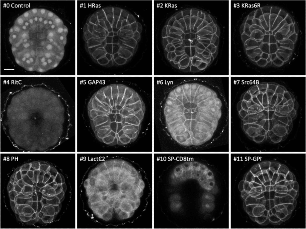
Targeting the cell membrane in established and emerging model organisms
Irene Karapidaki, Mette Handberg-Thorsager, Tsuyoshi Momose, Hitoyoshi Yasuo, Grigory Genikhovich, Sarah Assaf, Clara Deleau, Ying Pang, Clayton Pavlich, Beke Lohmann, Maria Lorenza Rusciano, Mattia Stranges, Juliette Mathieu, Marie Zilliox, Kirill Ustyantsev, Bastien Salmon, Béryl Laplace-Builhé, Manon Koenig, Jeffrey J. Colgren, Maria Ina Arnone, Eugene Berezikov, Thibaut Brunet, Gregor Bucher, Pawel Burkhardt, Daniel J. Dickinson, Evelyn Houliston, Jan Huisken, Lucas Leclère, Michalis Averof
ColorSTEM: An easy and convenient approach to Multicolor Electron Microscopy of Labeled Biological Specimens
Ranjan Ramachandra, Mason R. Mackey, Junru Hu, Tristan M. Shone, Mia V. Flores, Isabella A. Ramos, Steven T. Peltier, Stephen R. Adams, Mark H. Ellisman
Self-blinking dye restores efficient use of nanobodies in single-molecule localization microscopy
Samrat Basak, Kaushik Inamdar, Yoav G. Pollack, László Albert, Daniel C. Jans, Stefan Jakobs, Jörg Enderlein, Roman Tsukanov, Felipe Opazo
Chiral Single Molecule Localization Microscopy (chiralSMLM)
Aravinth S, Neeraj Pant, Partha Pratim Mondal
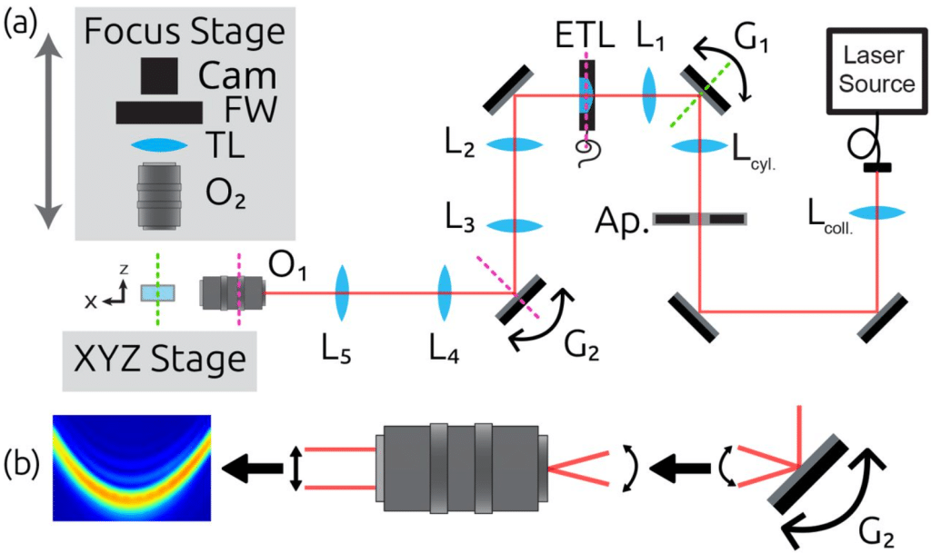
Curved Axially Scanned Light-Sheet Microscopy
Steven J. Sheppard, Tatsuya C. Murakami, Douglas P. Shepherd
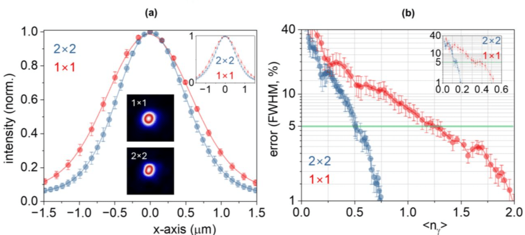
Quantitative STED microscopy with DNA-fluorophore labels
Laurell F. Kessler, Yunqing Li, Ashwin Balakrishnan, Mike Heilemann
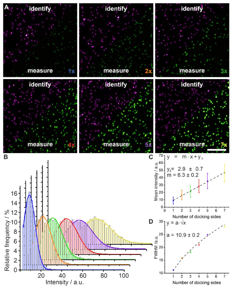
Figure extracted from Kessler, et al. The image is made available under a CC-BY 4.0 International license.
Unraveling Subcellular Ultrastructure with Cyclically Multiplexed Expansion Microscopy
Seweryn Gałecki, Bo-Jui Chang, Felix Zhou, Qionghua Shen, Daniel Stoddard, Bingying Chen, Daniela Nicastro, Reto Fiolka, Kevin M. Dean
Ångström Resolution with Flow Immunofluorescence Localization Microscopy (FILM)
Johanna Bartmuß, Anke Leinhaas, Tatjana Frank-Wiebe, Marc Schmidt-Supprian, Ali Kinkhabwala
Residual blinking-driven channel alignment for multicolor single-molecule localization microscopy
Fen Hu, Jianyu Yang, Mingxin Chen, Zhao Xie, Shuai Liu, Dan Ding, Leiting Pan, Jingjun Xu
Enhancing the imaging rate of high-speed atomic force microscopy using a combination of multiple techniques
Shingo Fukuda, Akihiro Otomo, Ryota Iino, Toshio Ando
Versatile high-speed volumetric imaging from microscopic to macroscopic scale by self-adaptive oblique plane microscopy
Dominique Meyer, Grant Kroeschell, Xiankun Lu, Linh Hoang, Haochen Wang, Shuying Li, Yu Kang T. Xu, Lei Tian, Jeff S. Mumm, Dwight E. Bergles, Ji Yi
Design and Fabrication of Petri Dish Optimized for High-Frequency Acoustic Microscopy Using 3D Printing
Tuna Pesen, Beyza Ceren Eren, Bora Akgun
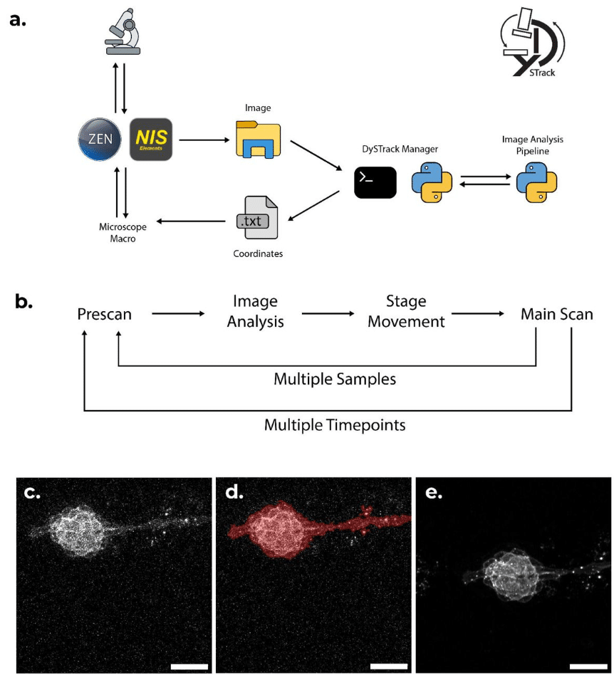
DySTrack: a modular smart microscopy tool for live tracking of dynamic samples on modern commercial microscopes
Zimeng Wu, Octavian Voiculescu, Alessandro Mongera, Roberto Mayor, Mie Wong, Jonas Hartmann
PKU tags, novel genetically encoded shape tags for cell labeling in light and electron microscopy
Rongbo Sun, Peilin Yang, Luyao Wang, Yixin Hu, Yini Yang, Fei Deng, Shaochuang Li, Mengyu Fan, Xiju Xia, Yulong Li
TransiScope: An Interactive Open-Source Platform for Automated Detection and Analysis of Transient Events in Time-Lapse Microscopy
Rinki Dasgupta, Kaushik Das
Enabling Real-Time Fluctuation-Based Super Resolution Imaging
Miyase Tekpinar, Jelle Komen, Hana Valenta, Ran Huo, Klarinda De Zwaan, Peter Dedecker, Nergis Tomen, Kristin Grussmayer
Spatial Polarization-Induced Fluorescence Fluctuation Imaging (SPIFFI) Enables Single-shot Super-Resolution and Multidimensional Imaging
Wei Guo, Lely Feletti, Aleksandra Radenovic
Pulsed-electron illumination does not reduce beam damage for imaging biological macromolecules
Vishal Kumar, Julika Radecke, K.V. Chinmaya, Inayathulla Mohammed, Ricardo C. Guerrero-Ferreira, Daniel Harder, Dimitrios Fotiadis, Henning Stahlberg
Mitigating the Field-of-View – Resolution Tradeoff by Photon Superlocalization
Sulaimon Balogun, Andreas E. Vasdekis
Photoejection turns non-covalent fluorescent tags into negative reversible photoswitchers
Yuriy Shpinov, Mrinal Mandal, Vincent van Deuren, Alienor Lahlou, Matthias Le Bec, Raja Chouket, Chaima Hadj Moussa, Clara Bonin, Hessam Sepasi Tehrani, Ian Coghill, Lina El Hajji, Karim Ounoughi, Jaime Franco Pinto, Marie-Aude Plamont, Philippe Pelupessy, Isabel Ayala, Franck Perez, Isabelle Aujard, Thomas Le Saux, Arnaud Gautier, Peter Dedecker, Bernhard Brutscher, Ludovic Jullien
Nanoscale imaging of native symbiotic animal tissue using a multimodal large volume imaging pipeline for cryo-electron tomography
Katrina A. Gundlach, Oda H. Schiøtz, Mark Ladinsky, Colin Raimann, Michael Rheinberger, Florian Beck, Barış Gündüz, Rob Langelaan, Martin Rücklin, Ronald W.A. Limpens, Edward G. Ruby, Margaret McFall-Ngai, Jürgen M. Plitzko, Ariane Briegel
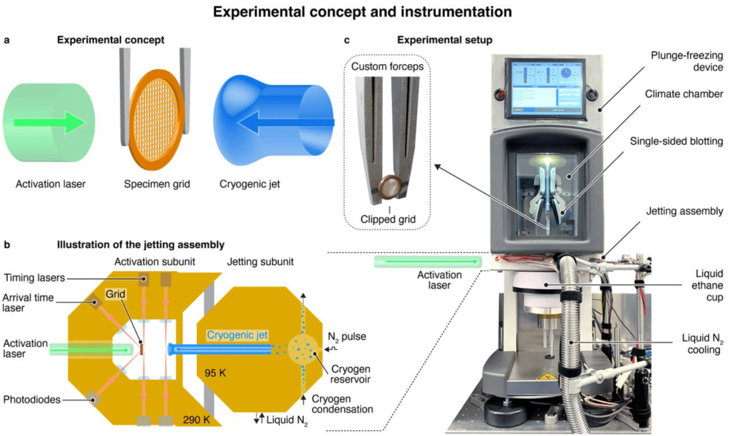
Microsecond Time-Resolved Cryo-EM Based on Jet Vitrification
Michal Haubner, Harry M. Williams, Jakub Hruby, Monique S. Straub, Albert Guskov, Kirill Kovalev, Marcel Drabbels, Ulrich J. Lorenz
Regioisomer-controlled red-shifted DNA probes for imaging of living tissues
Kamila A. Kiszka, Shalini Pradhan, Jonas Bucevičius, Tanja Koenen, Gražvydas Lukinavičius
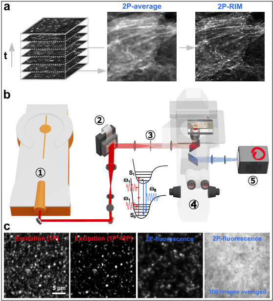
Widefield two-photon random illumination microscopy (2P-RIM)
Assia Benachir, Xiangyi Li, Eric M. Fantuzzi, Guillaume Giroussens, Thomas Mangeat, Federico Vernuccio, Hervé Rigneault, Anne Sentenac, Sandro Heuke
Nanodiamond-Enabled Torsion Microscopy Uncovers Multidimensional Cell-Matrix Mechanical Interactions
Yong Hou, Lingzhi Wang, Zheng Hao, Fuqiang Sun, Yutong Wu, Luyao Zhang, Linjie Ma, Wenyan Xie, Xinhao Hu, Qiang Wei, Cheng-han Yu, Yuan Lin, Zhiqin Chu
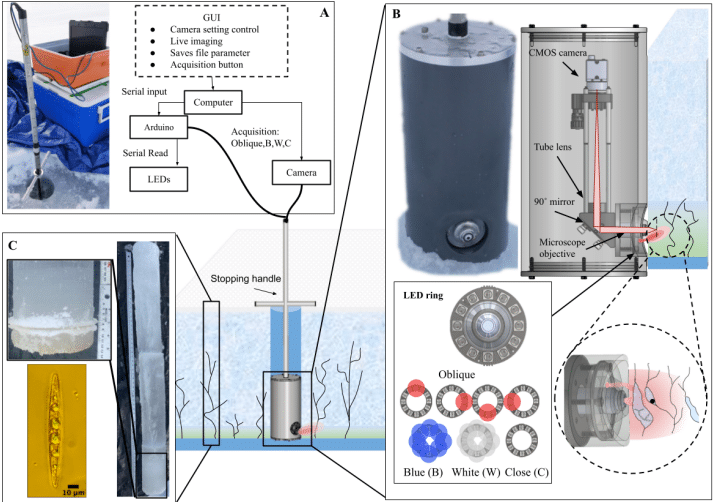
Microscopy system for in situ sea ice structure and biology observations
Lessard-Hamel Béatrice, Babin Marcel, Thibault Simon
High Space-bandwidth Product Label-free Examination of iPSC-derived Brain Organoids via Fourier Ptychographic Microscopy
Mikolaj Krysa, Mikolaj Rogalski, Piotr Arcab, Pawel Goclowski, Kamil Kalinowski, Piotr Zdańkowski, Vishesh K. Dubey, Mukesh Varshney, Balpreet S. Ahluwalia, Maciej Trusiak
Restless Multi-Process Multi-Armed Bandits with Applications to Self-Driving Microscopies
Jaume Anguera Peris, Songtao Cheng, Hanzhao Zhang, Wei Ouyang, Joakim Jaldén
Quantum-optimal nonlinear microscopy with classical light
Joshua L. Reynolds, Shaun C. Burd, Tzu-Chieh Yen, Samsuzzoha Mondal, Soichi Wakatsuki, Mark A. Kasevich
Real-Time Control and Automation Framework for Acousto-Holographic Microscopy
Hasan Berkay Abdioğlu, Yağmur Işık, Mustafa İsmail İnal, Nehir Serin, Kerem Bayer, Muhammed Furkan Koşar, Taha Ünal, Hüseyin Üvet
Self-supervised prior learning improves structured illumination microscopy resolution
Ze-Hao Wang, Tong-Tian Weng, Long-Kun Shan, Xiang-Dong Chen, Guang-Can Guo, Fang-Wen Sun, Tian-Long Chen
Depth-enhanced molecular imaging with two-photon oblique plane microscopy
Kevin Keomanee-Dizon, Yaakov Clenman, Alejandra Duran, Sergey Ryabichko, Pauline Hansen, Tohn Borjigin, Richard Thornton, Jared E. Toettcher, Harold M. McNamara
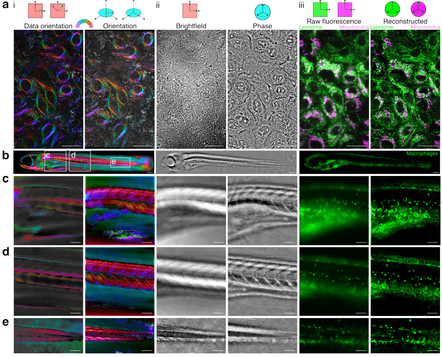
WaveOrder: A differentiable wave-optical framework for scalable biological microscopy with diverse modalities
Talon Chandler, Ivan E. Ivanov, Gabriel Sturm, Sheng Xiao, Xiang Zhao, Alexander Hillsley, Allyson Quinn Ryan, Ziwen Liu, Sricharan Reddy Varra, Ilan Theodoro, Eduardo Hirata-Miyasaki, Deepika Sundarraman, Amitabh Verma, Madhurya Sekhar, Chad Liu, Soorya Pradeep, See-Chi Lee, Shannon N. Rhoads, Maria Clara Zanellati, Sarah Cohen, Carolina Arias, Manuel D. Leonetti, Adrian Jacobo, Keir Balla, Loïc A. Royer, Shalin B. Mehta
 (No Ratings Yet)