Vote for the cover – #JCSImagingSI
Posted by FocalPlane, on 25 September 2024
Thanks to everyone who entered our image competition to select the cover for the Journal of Cell Science Special Issue: Imaging Cell Architecture and Dynamics. With so many fantastic images to choose from our Executive Editor Seema Grewal, and Guest Editors Lucy Collinson (The Francis Crick Institute, UK) and Guillaume Jacquemet (University of Turku, Finland) have shortlisted 10 images for a public vote.
Vote for your favourite image using the poll at the bottom of the page. The winning image will be featured on the cover of JCS and the winning scientist will receive £200. The special issue is already open and we’ll be adding more articles until the issue closes in October. You can check out the table of contents here.
Voting closes on Wednesday 9 October at 16:00 BST.
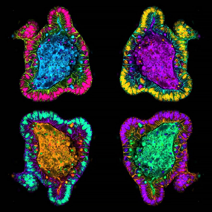
1. Myo5b KO organoids – Amy Engevik
Intestinal organoids represent an in vitro system that closely recapitulates in vivo cell biology and physiology. These intestinal organoids were generated from mice lacking Myosin 5b which results in cellular abnormalities including apical membrane proteins being retained subapically. This micrograph was acquired on a Zeiss Axiocam microscope.
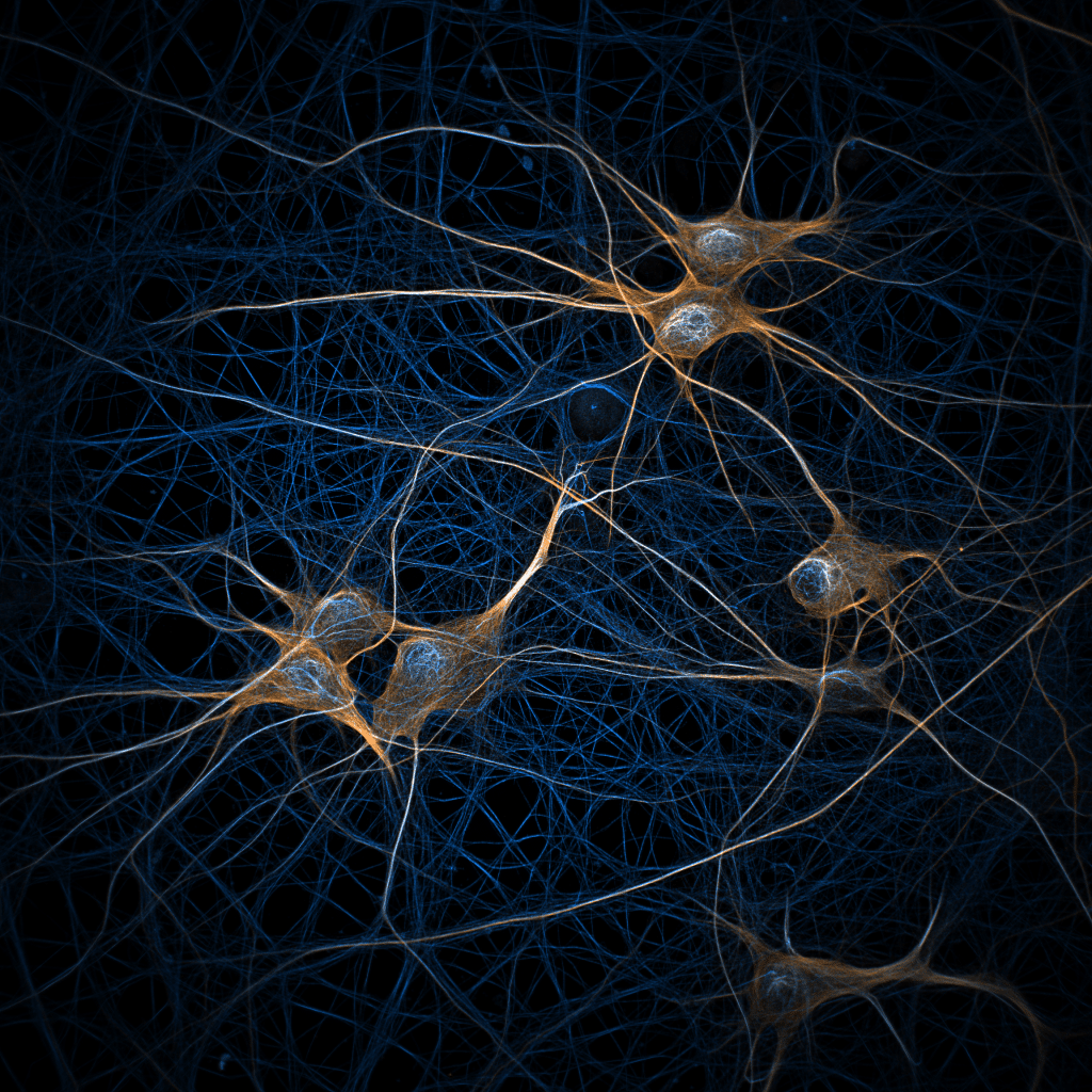
2. Neuronal Space Invaders – Ciarán Butler-Hallissey
These rat hippocampal neurons are labelled to show microtubules in blue and MAP2 in orange. The image was acquired on spinning disk confocal microscope.
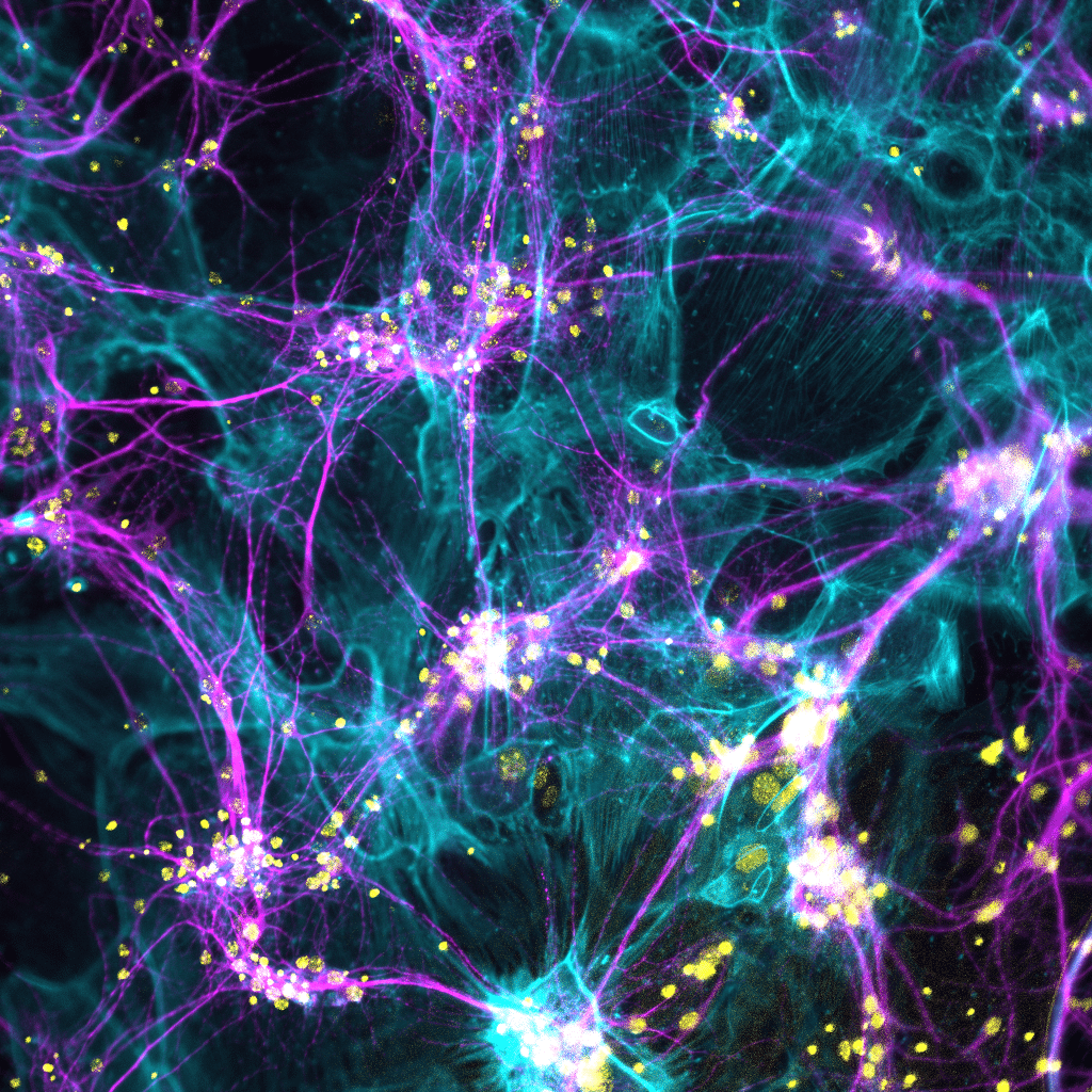
3. Neuronal galaxy – Mari Angeles Juanes
Mature neurons were fixed and stained with MAP2 antibody (neurite marker, cyan), Alexa Fluor 488-phalloidin to visualize F-actin, and DAPI (nuclei, yellow). The image shows the connections between the actin and microtubule cytoskeletons. The images was acquired at 20x on a widefield Leica DMi8 microscope, merged and colour-coded in Fiji.
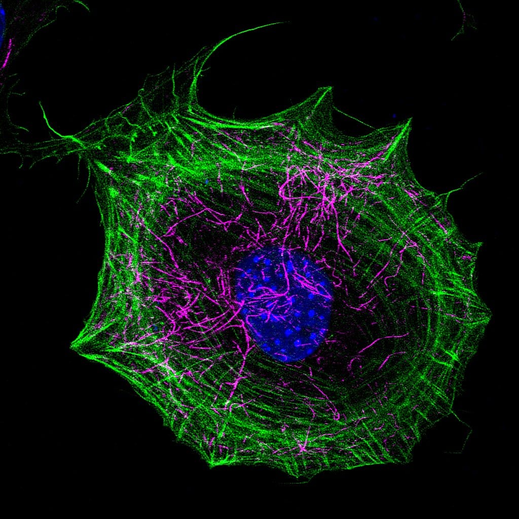
4. Breaking Symmetry – Antara Chakraborty
WT-MEF labelled with phalloidin (actin – green), acetylated tubulin antibody (magenta) and DAPI (DNA – blue). The image was captured using Laser scanning confocal microscope 780. Deconvolution was done on Huygens Professional version 16.10 and the image was reconstructed using ImageJ.
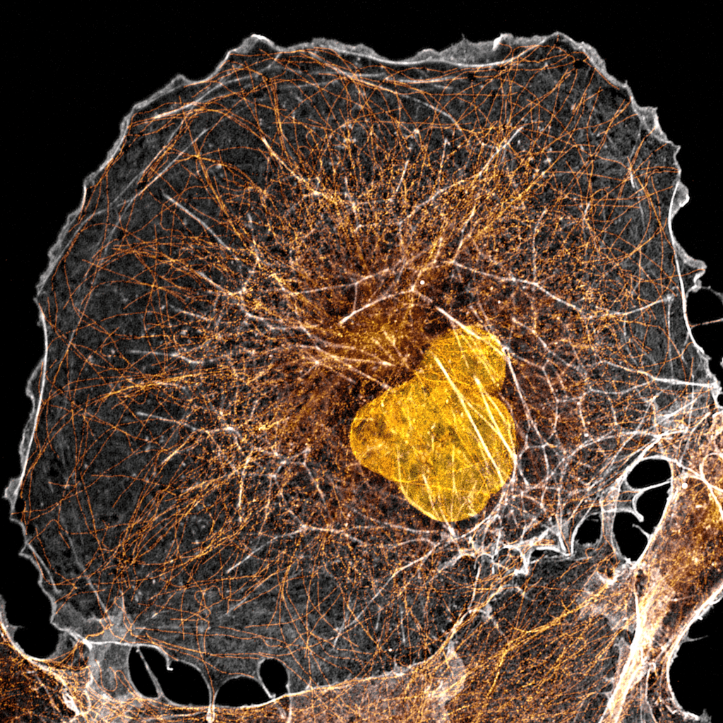
5. Cell Cytoskeleton – Joe McKellar
This image depicts an African Green Monkey cell stained for actin (white), microtubules (gold) and DNA (orange). The image was taken with an LSM 980 confocal microscope with Airyscan 2 module in Airyscan mode using a 63x lens. Airyscan processing was done on ZEN Blue and post-processing was done on Fiji.
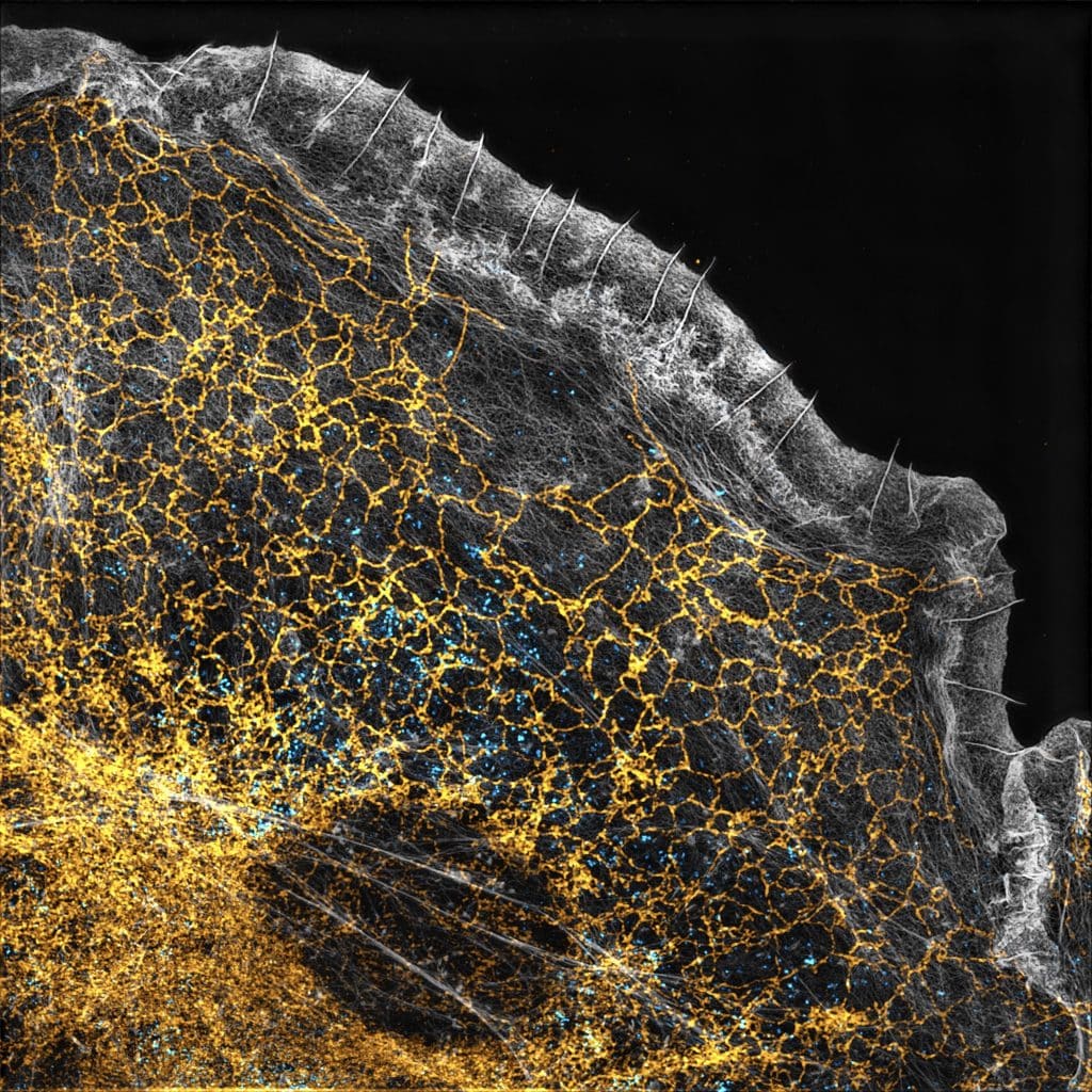
6. Rising tide – Christophe Leterrier
COS-7 cell labeled for actin (phalloidin, gray), ER (RTN4, yellow) and clathrin (cyan) imaged by 3D-SIM, processed with Fiji.
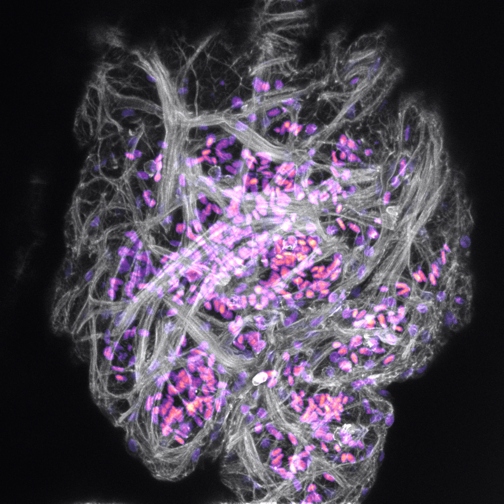
7. Dynamic atrial architecture – Aaron Scott
The myocardial actin networks of the adult zebrafish atrial chamber, bulging with blood cells. The heart was fixed and stained to show nuclei (hot purple) and F-actin networks (grey), and then mounted in agarose before imaging. The image was acquired using Leica SP8 AOBS confocal laser scanning microscope and reconstructed in ImageJ.
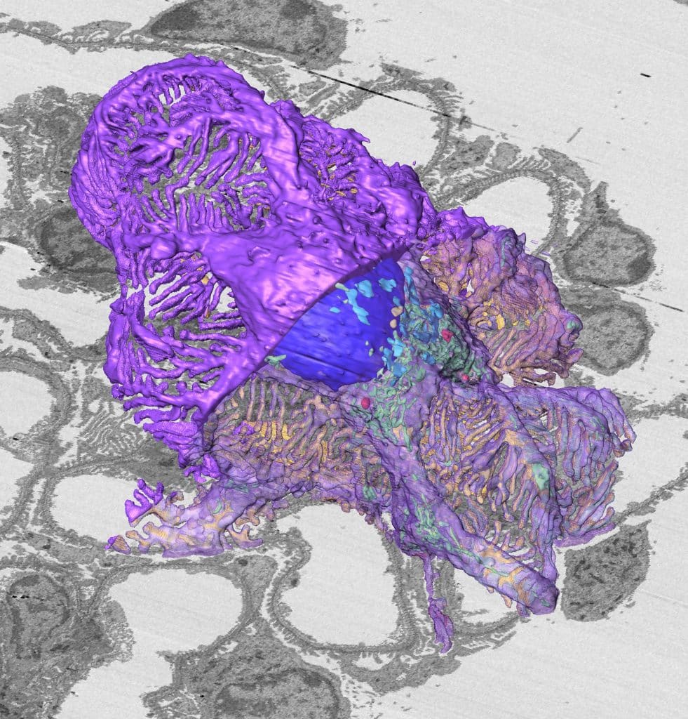
8. Mouse Podocyte – Takayuki Miyaki
3D ultrastructural reconstruction of a single mouse podocyte. Podocytes are specialized epithelial cells for blood filtration in the kidney glomeruli. This reconstruction, performed using Amira, is based on complete serial sectional images of a whole glomerulus obtained using field-emission electron microscopy (FE-SEM). The podocyte’s luminal cell membrane (purple) is partially translucent, indicating the localization of the organelle (yellow, centrosome; red, lysosomes; green, mitochondria; dark blue, nucleus; light blue, Golgi apparatuses). The yellow membrane is the basal cell membrane.
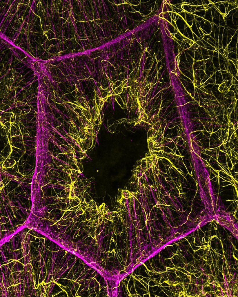
9. Intricate Interactions: The Dance of Microtubules and Actin – Wen Lu
The image displays the intricate internal cytoskeletal structure of a nurse cell within a Drosophila ovary. Microtubules were labelled with EMTB-3XGFP (yellow), and actin filaments were stained with Rhodamine-conjugated phalloidin (magenta). The image is a maximum intensity projection of a Z-stack image, acquired on DeepSIM by CrestOptics, and it was processed in Fiji.
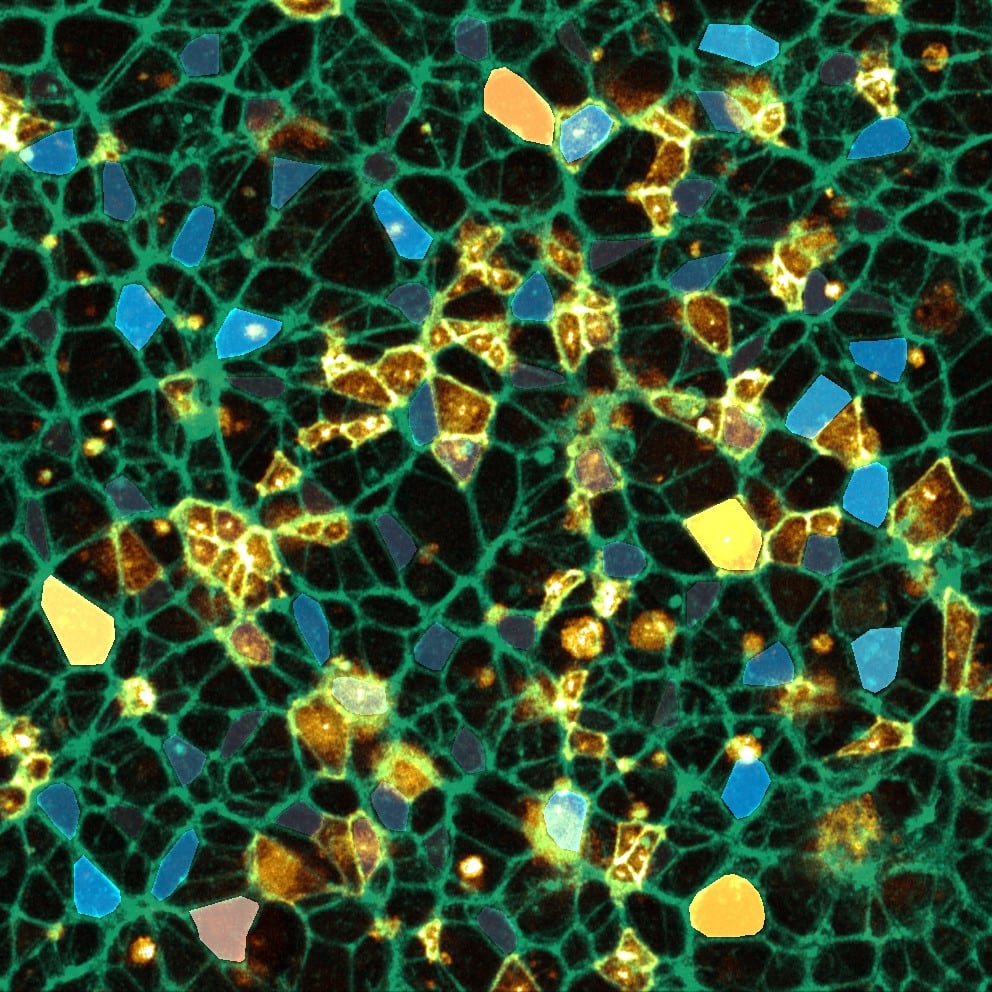
10. Pebbles – Ioakeim (Makis) Ampartzidis
In this image, the actin cytoskeleton of neuroepithelial cells is shown in green, and the membranes of some randomly selected cells are highlighted in yellow using synthetic fluorescent labelling dyes. The apical membranes of some cells were pseudo-coloured based on their size, ranging from light blue (small size) to orange (large apical area). The image was taken with the ZEISS LSM880 upright microscope with Airyscan imaging and processed in ImageJ.
Don’t forget to check out the Journal of Cell Science Special Issue: Imaging Cell Architecture and Dynamics.
Voting closes on Wednesday 9 October at 16:00 BST.
Thank you for voting


 (39 votes, average: 1.00 out of 1)
Clear image
I love this image
Wow. beautiful pictures
It is fantastic.
Good
😍
Good Work
voting for number 2 space invaders;
all stunning images but this is my favourite for its clarity and overall composition. I also like the title, with the image clearly demonstrating the idea 😉
Clear and mesmerizing
Very good image captured showing the asymmetry
Everything is so so beautiful! It was difficult to vote!
My vote is for image 4
The pictures are all sharp and clear, had a tough time to select the best
Amazing
Awesomly detailed image
Wonderful connections, sharp and shinny;)
Great learning here
Gorgeous Neurons #3!
Gorgeous pic #3
Fantastic
No. 9 is such an impressive image. Voting for #9.
I am voting for #8. LIght in color, attractive.
Lovely images! All would look great on the cover – best of luck to all the entrants
Vote for no. 4
Very interesting images with proper symmetry around
#9 is awesome! Love the dance of yellow and magenta!
All beautiful images!
Personally I will vote for #9. Love the color combination.
#2 is stunning