Microscopy preprints – new tools and techniques in imaging
Posted by FocalPlane, on 13 December 2024
Here is a curated selection of preprints posted recently on new tools and techniques in imaging. Let us know if we are missing any recent preprints that are on your reading list!
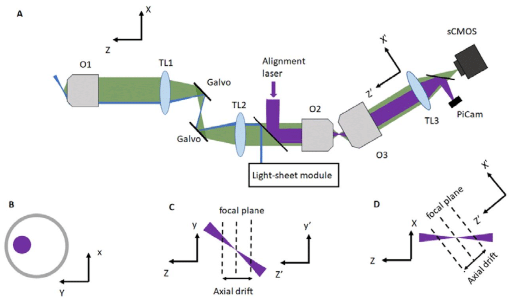
Active Remote Focus Stabilization in Oblique Plane Microscopy
Trung Duc Nguyen, Amir Rahmani, Aleks Ponjavic, Alfred Millett-Sikking, Reto Fiolka
Multiplexed Ultrasound Imaging of Gene Expression
Nivin N. Nyström, Zhiyang Jin, Marisa E. Bennett, Ruby Zhang, Margaret B. Swift, Mikhail G. Shapiro
Integrating Machine Learning with Flow-Imaging Microscopy for Automated Monitoring of Algal Blooms
Farhan Khan, Benjamin Gincley, Andrea Busch, Dienye L Tolofari, John W Norton Jr, Emily Varga, R Michael Mckay, Miguel Fuentes-Cabrera, Tad Slawecki, Ameet J. Pinto
All-Optical Multimodal Mapping of Single Cell Type-Specific Metabolic Activities via REDCAT
Yajuan Li, Zhaojun Zhang, Archibald Enninful, Jungmin Nam, Xiaoyu Qin, Jorge Villazon, Negin Fazard, Anthony A. Fung, Mina L. Xu, Hongje Jang, Nancy R. Zhang, Rong Fan, Zongming Ma, Lingyan Shi
A toolkit for testing membrane localisation tags across species
Irene Karapidaki, Tsuyoshi Momose, Marie Zilliox, Michalis Averof
A green lifetime biosensor for calcium that remains bright over its full dynamic range
Franka H. van der Linden, Stephen C. Thornquist, Rick M. ter Beek, Jelle Y. Huijts, Mark A. Hink, Theodorus W.J. Gadella Jr., Gaby Maimon, Joachim Goedhart

Calibration-free estimation of field dependent aberrations for single molecule localization microscopy across large fields of view
Bernd Rieger, Sjoerd Stallinga, Isabel Droste, Keith A Lidke, Sajjad Khan, Ben van Werkhoven, Stijn Heldens, Erik Schuitema
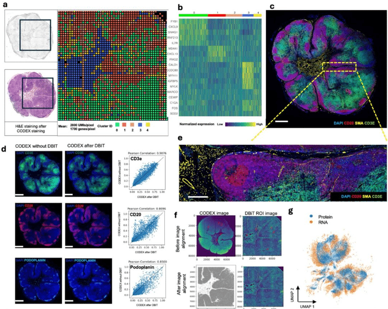
Integration of Imaging-based and Sequencing-based Spatial Omics Mapping on the Same Tissue Section via DBiTplus
Archibald Enninful, Zhaojun Zhang, Dmytro Klymyshyn, Hailing Zong, Zhiliang Bai, Negin Farzad, Graham Su, Alev Baysoy, Jungmin Nam, Mingyu Yang, Yao Lu, Nancy R. Zhang, Oliver Braubach, Mina L. Xu, Zongming Ma, Rong Fan
Histology-Guided Single-Cell Mass Spectrometry Imaging using Integrated Bright-field and Fluorescence Microscopy
Alexander Potthoff, Marcel Niehaus, Sebastian Bessler, Jan Schwenzfeier, Emily Hoffmann, Oliver Soehnlein, Jens Höhndorf, Klaus Dreisewerd, Jens Soltwisch
Semi-automated navigation for efficient targeting of electron tomography to regions of interest in volume correlative light and electron microscopy
Kohki Konishi, Guilherme Neves, Matthew Russell, Masafumi Mimura, Juan Burrone, Roland Fleck
Fast Photostable Expansion Microscopy Using QDots and Deconvolution
Loku Gunawardhana, Wilna Moree, Jiaming Guo, Camille Artur, Tasha Womack, Jason L. Eriksen, David Mayerich
Monitoring the Coating of Single DNA Origami Nanostructures with a Molecular Fluorescence Lifetime Sensor
Michael Scheckenbach, Gereon Andreas Brüggenthies, Tim Schröder, Karina Betuker, Lea Wassermann, Philip Tinnefeld, Amelie Heuer-Jungemann, Viktorija Glembockyte
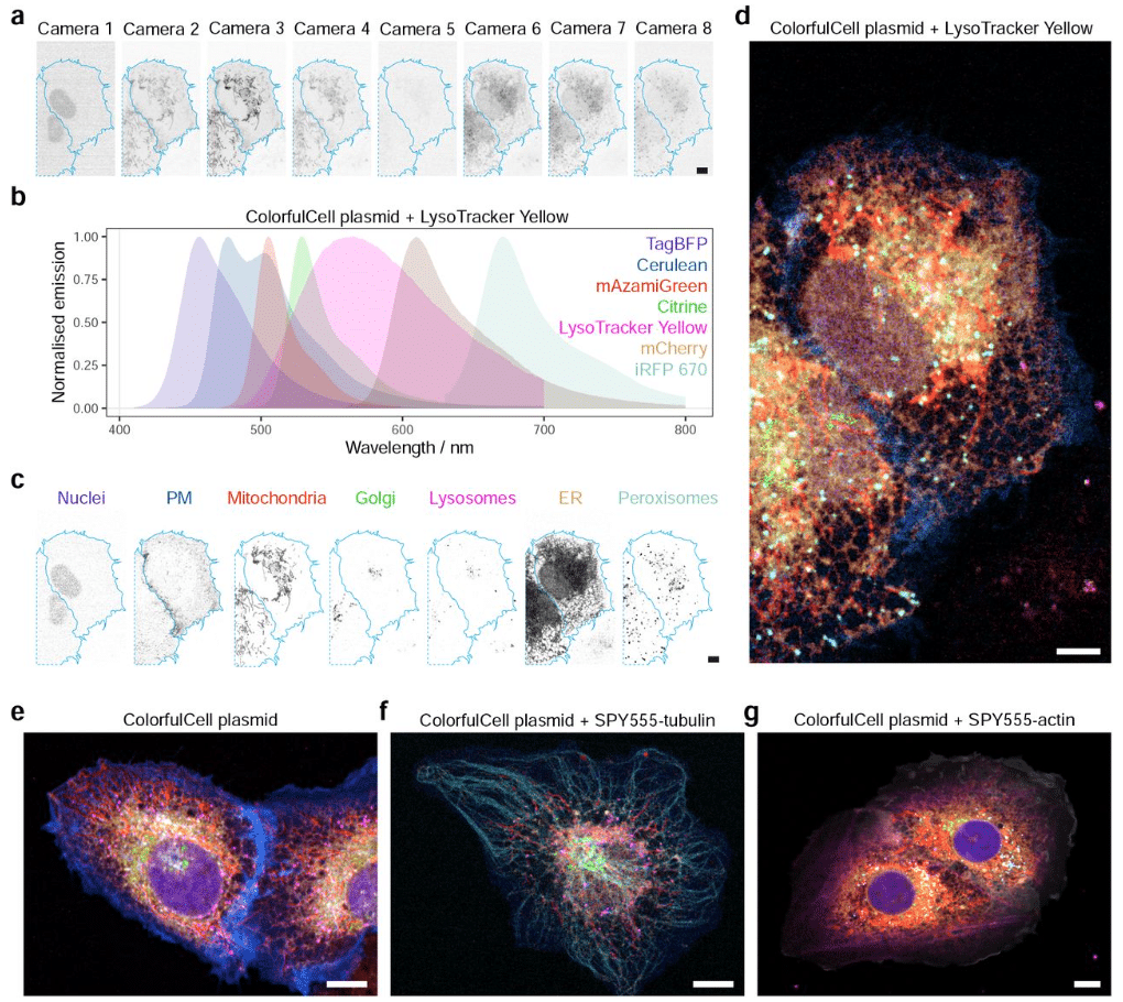
Multispectral live-cell imaging with uncompromised spatiotemporal resolution
Akaash Kumar, Kerrie E. McNally, Yuexuan Zhang, Alex Haslett-Saunders, Xinru Wang, Jordi Guillem-Marti, David Lee, Buwei Huang, Sjoerd Stallinga, Robert R. Kay, David Baker, Emmanuel Derivery, James D. Manton
Adaptive optical correction for in vivo two-photon fluorescence microscopy with neural fields
Iksung Kang, Hyeonggeon Kim, Ryan Natan, Qinrong Zhang, Stella X. Yu, Na Ji
Multiscale Light Field Microscopy Platform for Multi-purpose Dynamic Volumetric Bioimaging
Yangyang Bai, Matt Jones, Lauro Sebastian Ojeda, Janielle Cuala, Lynne Cherchia, Senta K. Georgia, Scott E. Fraser, Thai V. Truong
High-throughput single molecule microscopy with adaptable spatial resolution using exchangeable oligonucleotide labels
Klarinda de Zwaan, Ran Huo, Myron N.F. Hensgens, Hannah Lena Wienecke, Miyase Tekpinar, Hylkje Geertsema, Kristin Grussmayer
The TriScan: fast and sensitive 3D confocal fluorescence imaging using a simple optical design
Robin Van den Eynde, Jon Verheyen, Paul Miclea, Josef Lazar, Wim Vandenberg, Peter Dedecker
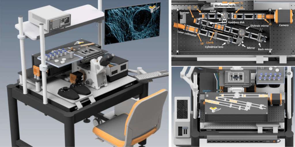
PISA: versatile microscope for 3D single molecule light sheet imaging
Yuichi Taniguchi, Kazuya Nishimura, Yamato Yoshida, Sooyeon Kim, Latiefa Kamarulzaman, David G. Priest, Masae Ohno
Optical Interference for the Guidance of Cryogenic Focused Ion Beam Milling Beyond the Axial Diffraction Limit
Anthony V. Sica, Magda Zaoralová, Cali Antolini, Daan B. Boltje, Judit J. Penzes, Lilyana M. Malmqvist, Grant Jensen, Jason T. Kaelber, Peter Dahlberg
Optical tomography reconstructing 3D motion and structure of multiple-scattering samples under rotational actuation
Simon Moser, Mia Kvåle løvmo, Franziska Strasser, Judith Hagenbuchner, Michael J. Ausserlechner, Monika Ritsch-Marte
Passage through micro-sprayer increases functional activity – implications for activity assays in time-resolved cryo-EM
Priyanka Garg, Xiangsong Feng, Swastik De, Joachim Frank
Up sampled PSF enables high accuracy 3D super-resolution imaging with sparse sampling rate
Jianwei Chen, Wei Shi, Jianzheng Feng, Jianlin Wang, Sheng Liu, Yiming Li
Impact of Triplet State Population on GFP-type Fluorescence and Photobleaching
Martin Byrdin, Svetlana Byrdina
A tunable and versatile chemogenetic near infrared fluorescent reporter
Lina El Hajji, Benjamin Bunel, Octave Joliot, Chenge Li, Alison G. Tebo, Christine Rampon, Michel Volovitch, Evelyne Fischer, Nicolas Pietrancosta, Franck Perez, Xavier Morin, Sophie Vriz, Arnaud Gautier
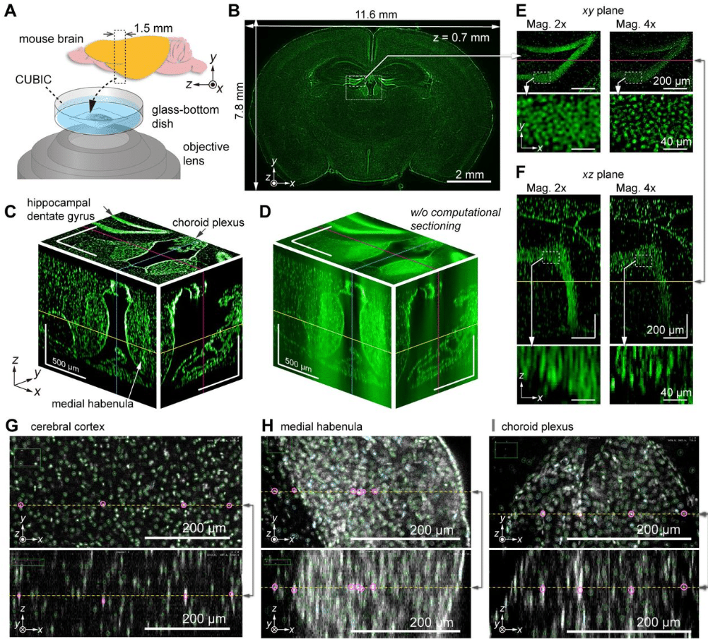
Volumetric trans-scale imaging of massive quantity of heterogeneous cell populations in centimeter-wide tissue and embryo
Taro Ichimura, Taishi Kakizuka, Yoshitsugu Taniguchi, Satoshi Ejima, Yuki Sato, Keiko Itano, Kaoru Seiriki, Hitoshi Hashimoto, Ko Sugawara, Hiroya Itoga, Shuichi Onami, Takeharu Nagai
Time-resolved cryo-EM (TRCEM) sample preparation using a PDMS-based microfluidic chip assembly
Xiangsong Feng, Joachim Frank
High SNR 3D Imaging from Millimeter-scale Thick Tissues to Cellular Dynamics via Structured Illumination Microscopy
Mengrui Wang, Manming Shu, Jiajing Yan, Chang Liu, Xiangda Fu, Jingxiang Zhang, Yuchen Lin, Hu Zhao, Yuwei Huang, Dingbang Ma, Yifan Ge, Huiwen Hao, Tianyu Zhao, Yansheng Liang, Shaowei Wang, Ming Lei
Experimental demonstration of Tessellation Structured Illumination Microscopy
Doron Shterman, Guy Bartal
Semicircular illumination scanning transmission electron microscopy
Akira Yasuhara, Fumio Hosokawa, Sadayuki Asaoka, Teppei Akiyama, Tomokazu Iyoda, Chikako Nakayama, Takumi Sannomiya


 (No Ratings Yet)