Featured image with Lea Berg
Posted by FocalPlane, on 4 July 2025
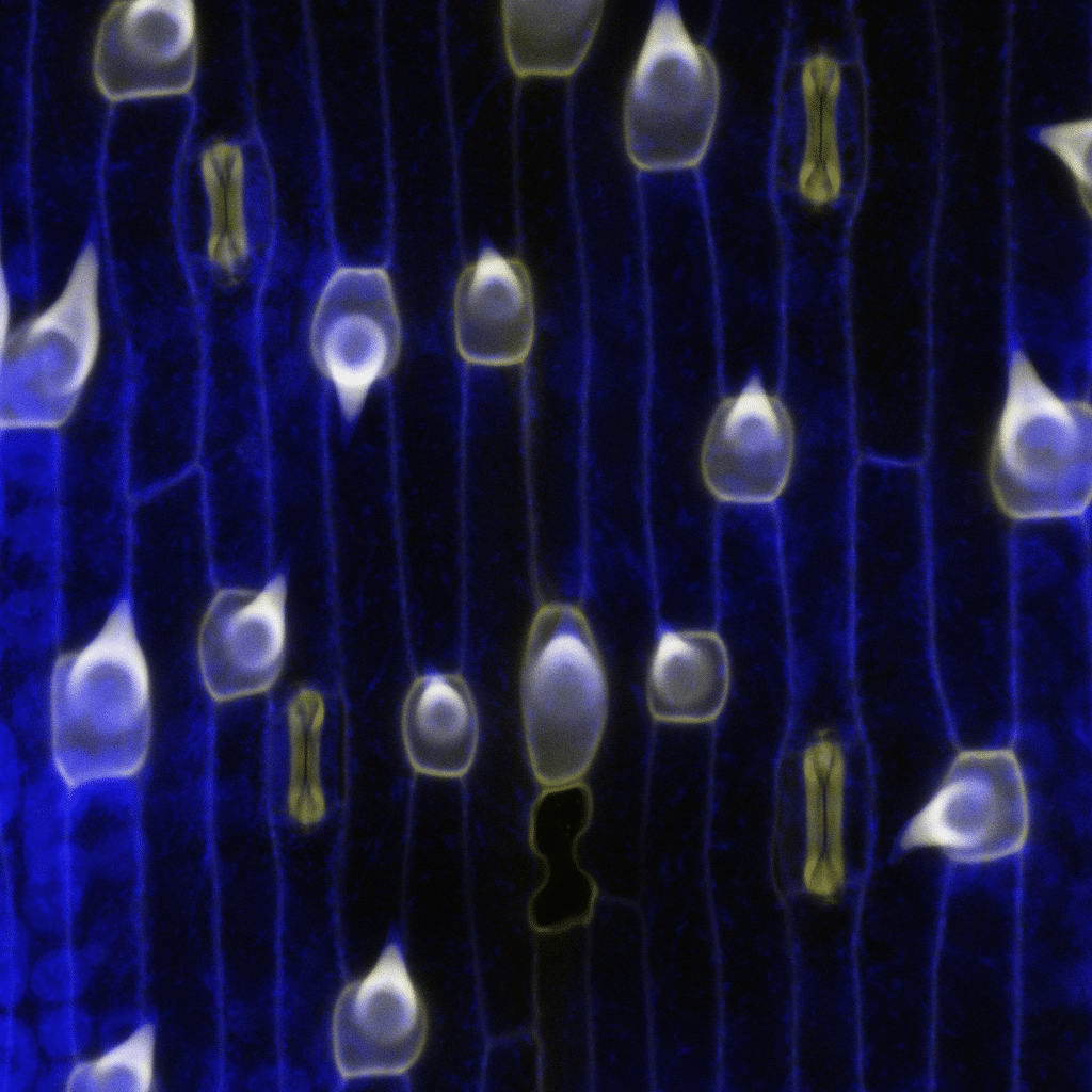
Our featured image, acquired by Lea Berg and Michael Rassig, shows mature epidermal cell types in a grass leaf of the emerging developmental model system Brachypodium distachyon. Cell outlines are blue, which is plant cell wall UV-autofluorescence. In yellow is stained lignin, a secondary cell wall modification that can be found in the hair cells (shark-tooth-shaped) and the stomatal guard cells (dumbbell-shaped). Imaged by confocal microscopy and processed in Fiji.
Discover more about Lea’s research
Research career so far: During my studies in Biological Sciences and Molecular Biosciences at the University of Heidelberg, I was attracted to the field of plant sciences where I was lucky to work on a variety of different species and topics. This enabled me to learn methods to study the physiology of plants, how to manipulate and explore their genetic background and how to image and analyze my samples using light and confocal microscopy. Towards the end of my studies in Heidelberg, I focused on grass stomatal biology which also lead me to my current research at the Stomatal Biology Lab at the Institute of Plant Sciences, University of Bern, Switzerland, where I am a fourth year PhD student.
Current research: My current research is focused on investigating the development of the early leaf epidermis using single-cell transcriptomics. I use various bioinformatic approaches to analyze my data in the search for potential new players in the early leaf stages. This then leads me back to the lab bench to verify gene expression using reporters and in situ hybridization, as well as analyzing and creating mutated plants to study the function of genetic candidates. My research is aimed at broadening our knowledge of these developmental processes so that we may better understand what makes the grasses such a successful plant family.
Favourite imaging technique/microscope: I enjoy confocal microscopy as it gives a very focused visualization and helps me to pinpoint where my signal can be found on a tissue and cellular level. The possibility of using different colours for the signal also makes it possible to create beautiful vibrant images and that is clearly a bonus on the artistic side. But I also like light microscopy as it allows me to quickly get an overview of my tissue of interest, which is very helpful when screening a lot of mutant plants.
What are you most excited about in microscopy? It is exciting to see how much further we can change and improve microscopy so that details and processes we could hardly see before can be visualized much better now and in the future. This will surely lead to new discoveries as well as increase the knowledge on already described processes in detail.
 (No Ratings Yet)