Featured image with Julia Zheku
Posted by FocalPlane, on 13 March 2026
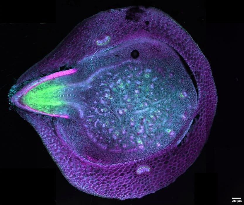
Our featured image, acquired by Julia Zheku, depicts a maize (corn) cross section, with a developing nodal root dynamically breaking through the basal stem node which is typically found below the soil surface. Such a root is not visible while sectioning, making this a beautiful, yet lucky, image. The section was prepared by mounting the sample in 5% agarose and then sectioning with a vibratome (Leica VT1000S). It was first fixed with a 4% formaldehyde fixative and then stained using Calcofluor White, Auramine O, and Direct Red 23. The sample was visualized using the Evident IXplore SpinSR Spinning Disk Confocal System and processed using ImageJ/Fiji and Canva.
Discover more about Julia’s research.
Research career so far: I began conducting research with Dr. Tobias Baskin on plant roots at the University of Massachusetts Amherst where I completed my undergraduate degree. Additionally, I also worked with Dr. Jaime Pinero on integrated pest management of early season insects on apple.
Current research: I recently have begun my master’s at the University of British Columbia (UBC), working with Dr. Arif Ashraf, to study primary root physiology in Arabidopsis, maize and wheat.
Favourite imaging technique: I enjoy using the spinning disk confocal. It is really user-friendly and customizable based on the kind of imaging you would like to do. The system that I am currently using at UBC’s bioimaging facility has two cameras, which provides beautiful high resolution images.
What are you most excited about in microscopy? I am really excited about vertical stage confocal microscopy. Specifically for live cell imaging of roots, it is extremely cool to see root cells dividing and elongating at the same time.
 (No Ratings Yet)