

Featured image with Mayuri Varma
Posted by FocalPlane, on 14 August 2026
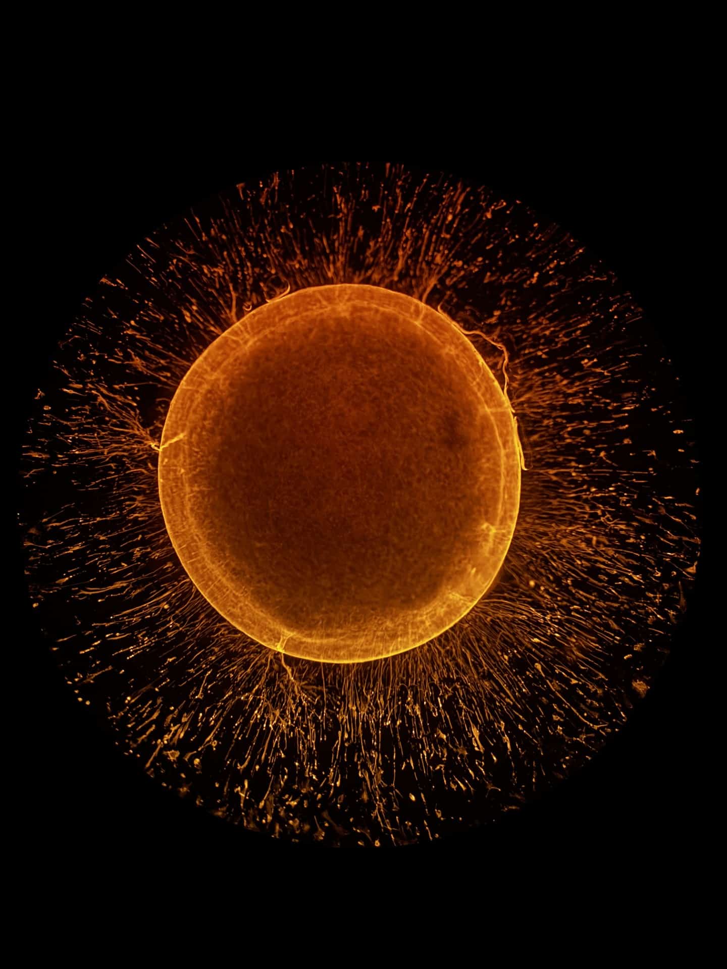
Our featured image, acquired by Mayuri Varma, shows a human iPSC-derived cortical organoid stained for glial fibrillary acidic protein (GFAP), a well-established marker of astrocytes. The organoid was generated from human induced pluripotent stem cells and immunostained to visualize astrocytic networks within the tissue. The striking radial organization of GFAP⁺ processes extending outward from a dense central core inspired the name “Astrocytic Supernova.” The sample was imaged using laser scanning confocal microscopy, and—perhaps most remarkably—the final image was captured simply by holding a phone camera to the ocular of the microscope, with no additional digital processing. What you see is exactly what was seen through the eyepiece.
Read more about Mayuri’s research.
Research career so far: I am a first-year PhD student in the stem cell field at the Centre for High Impact Neuroscience and Translational Applications (CHINTA), TCG CREST, India, and an AcSIR research scholar. Prior to my PhD, my research spanned two distinct but complementary areas—investigating the neuroprotective effects of natural compounds in a murine model of smokeless tobacco toxicity and exploring exosomal microRNAs as potential biomarkers for Parkinson’s disease. These experiences deepened my curiosity about disease mechanisms at the cellular level and ultimately drew me towards stem cell-based models of neurodegeneration.
Current research: My PhD research focuses on developing human iPSC-derived brain organoid models to study Alzheimer’s disease. I am particularly interested in glial biology—specifically the roles of astrocytes and microglia in neuroinflammation, protein homeostasis, and neuron–glia interactions. By leveraging these human-relevant models, I hope to uncover early disease mechanisms and identify potential therapeutic targets that may one day translate into meaningful clinical strategies.
Favourite imaging technique/microscope: Confocal laser scanning microscopy is one of my favorite imaging techniques. I find particular joy in reconstructing z-stacks, as it allows me to appreciate the intricate spatial organization of cells and their interactions within an intact, biologically relevant structure. There is something deeply satisfying about watching a three-dimensional world emerge from a stack of optical slices.
What are you most excited about in microscopy? I am most excited by the rapid advances in high-resolution and live-cell imaging that are enabling us to observe cellular behavior in real time within physiologically relevant models. When combined with human brain organoids and quantitative image analysis pipelines, these approaches hold enormous potential to reveal disease mechanisms that were simply impossible to study until recently. I believe we are entering an era where microscopy will not just document biology—it will actively drive discovery.