

Taking DNA-PAINT deeper: accessible super-resolution imaging in whole cells and tissues
Posted by sabrina.simoncelli, on 18 November 2026
by Cecilia Zaza and Sabrina Simoncelli
How can we visualise molecular organisation deeper within biological samples?
Single-molecule localisation microscopy (SMLM) allows us to investigate biological structures at the nanoscale, revealing details that conventional fluorescence microscopy cannot resolve. However, obtaining this level of detail deeper within cells and tissues remains challenging. Background fluorescence, limited photon collection and the need for optical sectioning can all affect image quality, particularly when we want to move beyond the thin region close to the coverslip. In practice, we are often forced to choose between resolution, imaging depth and field of view.
We wanted to explore how we could combine the high spatial resolution of DNA-PAINT with the optical capabilities of spinning-disk confocal microscopy with optical photon reassignment (SDC-OPR), making high-resolution imaging deeper within biological samples more accessible to researchers.
What does SoRa bring to DNA-PAINT?
DNA-PAINT uses transient binding between complementary DNA strands to generate the individual fluorescent events needed to localise molecules with nanometre precision1 (Fig. 1a). The high photon budget of individual binding events, together with the programmability of DNA interactions, makes DNA-PAINT a powerful approach for resolving nanoscale molecular organisation and enables multiplexed imaging of multiple targets through sequential rounds of imaging.
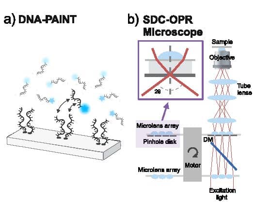
In our work, we combined DNA-PAINT with a Nikon Ti2 microscope equipped with the Yokogawa CSU-W1 SoRa spinning-disk confocal system2. The SoRa microscope incorporates a microlens array that enables optical photon reassignment, improving effective spatial resolution and photon collection (Fig. 1b). Combined with the optical sectioning provided by confocal microscopy, this creates an attractive platform for imaging samples in which out-of-focus fluorescence and photon collection become increasingly important.
Previous work has demonstrated the potential of conventional spinning-disk confocal microscopy for DNA-PAINT imaging in whole cells3. Our approach builds on this foundation by incorporating SoRa’s microlens-based optical photon reassignment. The microlens array positioned in front of the pinholes redistributes detected fluorescence towards the corresponding pinhole centres, allowing a greater proportion of emitted photons to contribute to image formation. This improves photon collection while retaining the optical sectioning benefits of confocal microscopy.
Importantly, our approach uses a commercially available microscope configuration rather than a bespoke optical system that researchers need to build and align themselves. SoRa systems are already available in many microscopy facilities worldwide, making this approach an attractive and accessible route for laboratories seeking to extend their super-resolution imaging capabilities to whole cells and tissues without developing a custom optical setup.
What did we discover when we moved from cells into tissues?
We first characterised the imaging performance using well-defined structures and cellular samples. In the basal plane, we demonstrated 6 nm spatial resolution, illustrating the level of structural detail achievable with the approach. We also showed that high localisation precision could be maintained at depth: in whole cells, we achieved sub-10 nm localisation precision at depths of up to 9 µm (Fig, 2a, b).
We then moved beyond cultured cells to investigate molecular organisation in tissue. Using the developing Drosophila retina, we imaged photoreceptor adherens junctions at depths of up to 15 µm, achieving an average localisation precision of 26 nm (Fig. 2c).
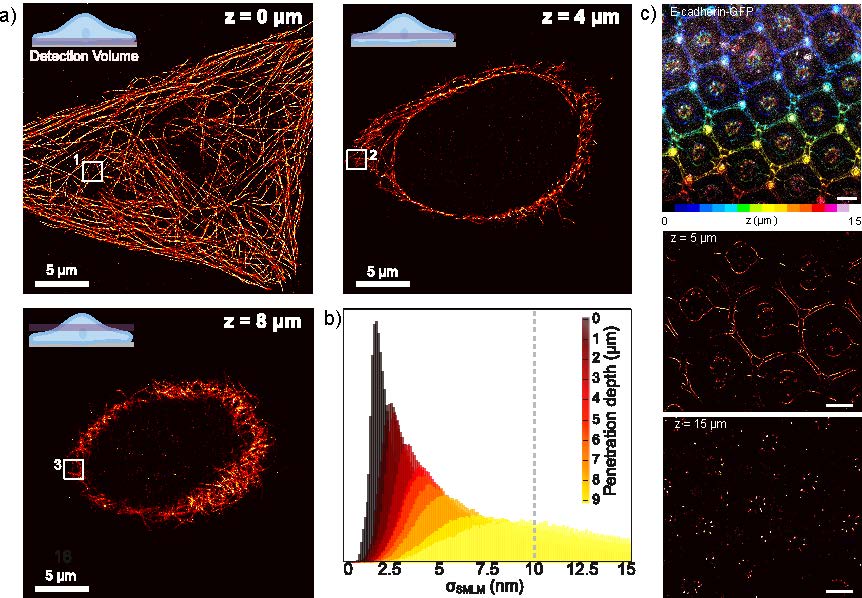
This was an especially exciting result because it demonstrated that the approach could be used to investigate nanoscale molecular organisation within a complex, developing tissue, rather than being restricted to regions close to the coverslip or isolated cells. It opens opportunities to study biological structures in their native spatial context, where molecular organisation can vary across cells, layers, and tissue regions.
Another interesting aspect of this experiment was that we could take advantage of existing GFP-expressing fly lines. By using commercially available DNA-conjugated anti-GFP nanobodies, we could directly label GFP-tagged proteins for DNA-PAINT imaging. This provides a convenient way to adapt existing fluorescent protein-tagged models to DNA-PAINT imaging, without the need to generate new constructs or develop custom DNA-conjugated antibodies.
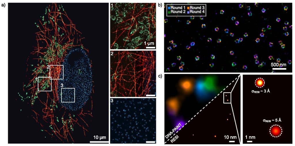
We also demonstrated that the system is compatible with the wider DNA-PAINT toolbox. Using Exchange-PAINT4 (Fig. 3a), we sequentially imaged microtubules, mitochondria and nuclear pore complexes in the same cell by exchanging the imager strands. We then applied RESI (Resolution Enhancement by Sequential Imaging)5 to resolve individual Nup96 proteins (Fig. 3c), achieving an average molecular precision of approximately 3 Å, representing a ten-fold improvement over a single round of DNA-PAINT imaging (Fig. 3c). Finally, we used quantitative DNA-PAINT (qPAINT)6 in Drosophila tissue to count molecules, measuring an average of 46 ± 27 collagen IV molecules per secretory vesicle.
How does this compare with other approaches to in-depth super-resolution imaging?
Several strategies have been developed to overcome the challenges of super-resolution imaging deeper within biological samples. These include approaches based on light-sheet illumination, which reduces background fluorescence and enables imaging across large sample volumes, as well as the integration of adaptive optics into super-resolution microscopy to compensate for sample-induced aberrations and improve image quality at depth. Each offers distinct advantages, with trade-offs in spatial resolution, photon collection, imaging depth, field of view and optical complexity.
Our approach offers a complementary route. By combining DNA-PAINT with the microlens-based optical photon reassignment of SoRa, we build on the optical sectioning capabilities of spinning-disk confocal microscopy to improve effective spatial resolution and photon collection. This enables high-resolution imaging across multiple cellular layers and within tissue samples, while retaining the practical advantages of a commercially available spinning-disk confocal platform.
The accessibility of instrumentation is particularly important. As SoRa is a commercially available optical configuration, researchers can benefit from microlens-based optical photon reassignment without having to develop, build and align a bespoke microscope. Although DNA-PAINT still requires appropriate sample preparation, labelling, acquisition and data analysis, our approach lowers the technical barrier to implementing in-depth single-molecule localisation microscopy.
Are there technical tips or tricks you have learnt, and/or technical challenges you had to overcome?
One of the main challenges when imaging deeper into biological samples was drift correction. Gold nanoparticles work well as fiducial markers close to the coverslip but become increasingly difficult to detect a few micrometres into the sample. At these depths we switched to a marker-free approach (AIM)7, which corrects drift using the DNA-PAINT localisations themselves.
Another challenge was maintaining sufficient photon collection with increasing imaging depth. Scattering and optical aberrations reduce the detected signal, which can compromise localisation precision. Maximising photon collection using SoRa, together with oxygen-scavenging imaging buffers (PCA/PCD/Trolox) to improve fluorophore performance, was therefore important.
Sample mounting also requires some optimisation. For the Drosophila eye discs, we modified a glass-bottomed dish so that the tissue layer of interest faced the objective, ensuring optimal optical access.
Finally, for 3D imaging, we generated an experimental point spread function (PSF) model from stacks of fluorescent beads, rather than introducing an astigmatic lens into the optical path. This meant we could perform 3D localisation without requiring additional optical components. Using this approach, we imaged mitochondria and resolved their characteristic hollow structure.
How can other laboratories use this method?
We wanted the approach to be useful beyond demonstrating what was possible in our own experiments. To support its adoption by other laboratories, we have also published a step-by-step protocol in the Journal of Visualized Experiments (JoVE)8, describing whole-cell super-resolution imaging via DNA-PAINT on a spinning-disk confocal microscope.
The protocol provides practical guidance through the entire experimental workflow, helping researchers implement the method and adapt it to their own samples. It covers buffer preparation, conjugation of antibodies to single-stranded DNA, preparation of the imaging buffer, microscope setup and optimisation, assessment of sample suitability for DNA-PAINT imaging, and finally, data processing and analysis to reconstruct super-resolution images.
What comes next?
We hope this approach will help researchers investigate molecular organisation in increasingly complex biological samples, extending single-molecule imaging beyond the coverslip-adjacent regions traditionally associated with TIRF-based SMLM.
Looking ahead, we see several exciting opportunities. Since SoRa systems are also used for live-cell imaging, the same microscope could potentially be used to follow biological processes in living cells and subsequently investigate their molecular organisation at the nanoscale using DNA-PAINT after fixation. Meanwhile, developments in faster and fluorogenic DNA-PAINT probes could help reduce background fluorescence and acquisition times, opening opportunities to image more challenging samples, including organoids and embryos. Combined with large fields of view and automated acquisition, these advances could eventually enable high-throughput nanoscale imaging across multiple cells and experimental conditions.
By combining the high photon budget of individual DNA-PAINT binding events with the improved photon collection provided by SoRa, we provide a practical route to high-resolution nanoscale imaging across whole cells and deeper within tissues. Ultimately, we hope this will encourage more researchers to explore molecular organisation in increasingly complex biological samples, bringing single-molecule imaging closer to the biological contexts in which these molecular structures actually function.
References
1. Schnitzbauer, J., Strauss, M. T., Schlichthaerle, T., Schueder, F. & Jungmann, R. Super-resolution microscopy with DNA-PAINT. Nat Protoc 12, 1198–1228 (2017).
2. Zaza, C. et al. Super-resolution imaging in whole cells and tissues via DNA-PAINT on a spinning disk confocal with optical photon reassignment. Nat Commun 16, 4991 (2025).
3. Schueder, F. et al. Multiplexed 3D super-resolution imaging of whole cells using spinning disk confocal microscopy and DNA-PAINT. Nat Commun 8, 2090 (2017).
4. Jungmann, R. et al. Multiplexed 3D cellular super-resolution imaging with DNA-PAINT and Exchange-PAINT. Nat Methods 11, 313–318 (2014).
5. Reinhardt, S. C. M. et al. Ångström-resolution fluorescence microscopy. Nature 617, 711–716 (2023).
6. Jungmann, R. et al. Quantitative super-resolution imaging with qPAINT. Nat Methods 13, 439–442 (2016).
7. Ma, H., Chen, M., Nguyen, P. & Liu, Y. Toward drift-free high-throughput nanoscopy through adaptive intersection maximization. Science Advances 10, eadm7765 (2024).
8. Zaza, C., Tanase, M., Dalby, O. P. L. & Simoncelli, S. Whole-cell Super-Resolution Imaging via DNA-PAINT on a Spinning Disk Confocal with Optical Photon Reassignment. Journal of Visualized Experiments (JoVE) e69531 (2026) doi:10.3791/69531.
Get involved
Create an account or log in to post your story on FocalPlane.
More posts like this

Filter by
- NewsApply
- DiscussionsApply
- How toApply
- ToolsApply
- Case studiesApply
- InterviewsApply
- JobsApply
- EducationApply
- Blog seriesApply
- Deep Learning for Bi..o-image analysisApply
- GloBIAS – updates fr..om the communityApply
- WAMBIAN: West Africa.. in FocusApply
- Volume EMApply
- Latin American Micro..scopistsApply
- Bio-image Analysis w..ith NapariApply
- Imaging with…Apply
- Towards Global Acces..sApply
- Latin America Bioima..gingApply
- From Zero to Qupath ..HeroApply
- Asian Microscopists ..and Cell BiologistsApply
- AIC at HHMI JaneliaApply
- Highlights from Euro..-BioImagingApply
- LSFM seriesApply
- DIY MicroscopyApply
- View all