Advertisement

Displaying posts with the tag: is_archive

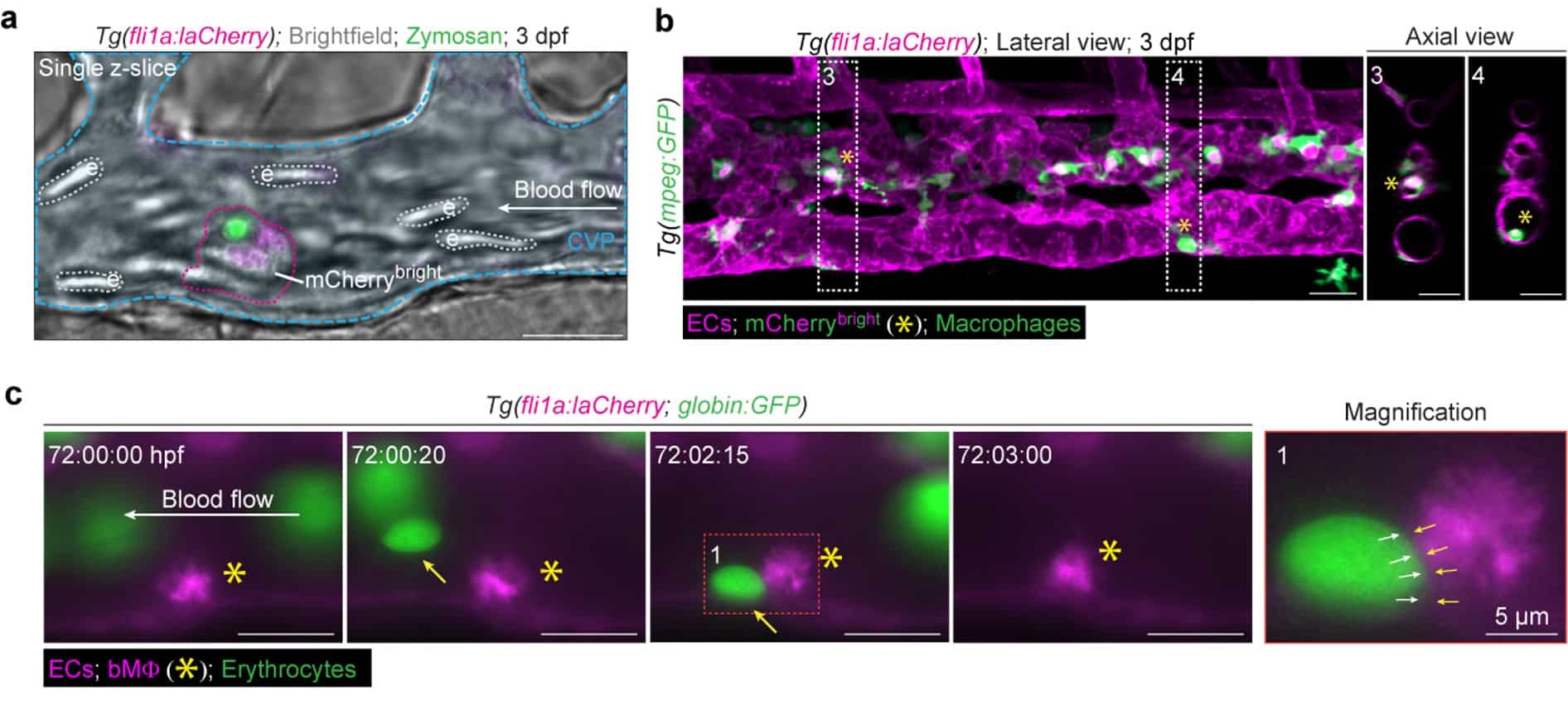
Seeing comes before knowing: how live imaging led to the discovery of blood vessel-resident macrophages

Posted by , on 12 August 2026

Bart Weijts and Catherine Robin share the behind the paper story on their discovery of blood vessel-resident macrophages.

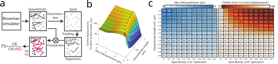
Imaging spotlight: FidlTrack

Posted by , on 10 August 2026

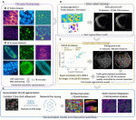
In this Imaging spotlight, we hear from Pierre Parutto and Edward Avezov about FidlTrack, a method for benchmarking and optimising single particle tracking.

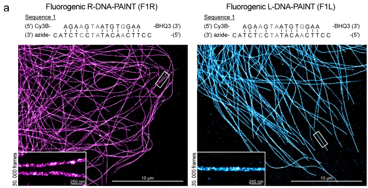
Imaging spotlight: fluorogenic L-DNA probes

Posted by , on 6 August 2026

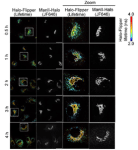
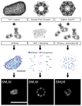
In this Imaging spotlight, we hear from Bas van Bommel and Helge Ewers about the fluorogenic L-DNA probes they used to optimise DNA-PAINT labelling for single molecule localisation microscopy.

Pearling and directed trafficking as determinants of mitochondrial genome organization and network architecture

Posted by , on 21 July 2026

In this highlight, we hear from Juan C. Landoni, who discusses his recent work uncovering how mitochondria distribute their genome through pearling (Landoni et al. 2026, Science), and the key role of mitochondrial trafficking in maintaining new material dissemination and network architecture (Winter, Landoni et al. 2026, JCB).

Compartmentalized Cytoplasmic Tradewinds

Posted by , on 9 July 2026

Cathy Galbraith and Jim Galbraith share their story of answering how does a cell deliver a soluble protein — untethered to any motor, riding on no track — to the right place at the right time? They used six different imaging approaches to discover hidden cytoplasmic compartments and tradewinds that were hiding in plain sight.

Imaging spotlight: VLab4Mic

Posted by , on 7 July 2026

In this preprint highlight, Damián Martínez, Bruno M. Saraiva, Mario Del Rosario, Ricardo Henriques introduce VLab4Mic, an open-source simulation platform that allows researchers to optimise microscopy experiments before performing them.

Imaging spotlight: Cryo-ExM reveals the architecture of the human cytotoxic immunological synapse

Posted by , on 15 June 2026

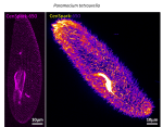
In this behind the paper story, Benita Wolf and colleagues share their research using cryo-expansion microscopy (cryo-ExM) to map the architecture of the human cytotoxic immunological synapse.

Imaging spotlight: PLIN5 phosphorylation tunes mitochondria–lipid droplet coupling in the liver

Posted by , on 29 April 2026

In this paper highlight, we hear from Natalie Porat-Shliom, who discusses her recent paper on how PLIN5 phosphorylation regulates mitochondria–lipid droplet contacts in the liver.

Imaging spotlight: Calibrating expansion microscopy to map receptor sorting domains in endosomes

Posted by , on 15 April 2026

In this behind the paper story, Tayla Shakespeare and Izzy Jayasinghe share their research mapping receptor sorting domains in endosomes using expansion microscopy (ExM). As part of this research, they developed a calibrated 3D ExM toolkit to measure local expansion.

Imaging spotlight: dissecting the precancerous niche

Posted by , on 25 March 2026

In this paper highlight we hear from Greta Skrupskelyte and Maria Alcolea, who discuss their recent paper dissecting the role of the microenvironment and tumour-stromal communication in tumour growth and survival.