Microscopy preprints – applications in cell biology and more
Posted by FocalPlane, on 8 April 2022
Here is a curated selection of preprints published recently. In this post, we focus specifically on preprints using microscopy tools in different fields such as cell biology, plant biology, microbiology, and neuroscience.
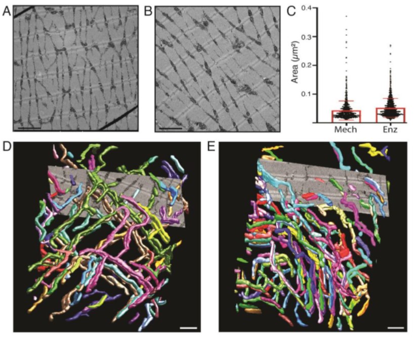
Justine M. Pinskey, Adhya Lagisetty, Long Gui, Nhan Phan, Evan Reetz, Gang Fu, Daniela Nicastro
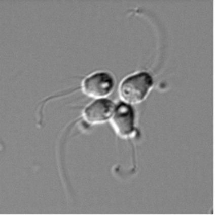
Kinetochore- and chromosome-driven transition of microtubules into bundles promotes spindle assembly
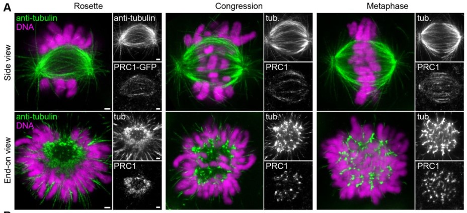
Jurica Matković, Subhadip Ghosh, Mateja Ćosić, Nenad Pavin, Iva M. Tolić
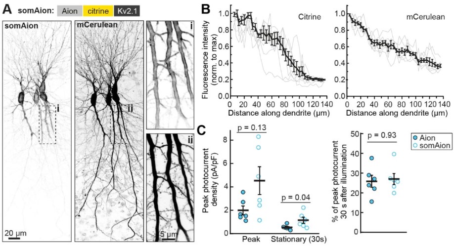
Temporally extended and reversible neuronal silencing with Aion
Silvia Rodriguez-Rozada, Jonas Wietek, Federico Marcello Tenedini, Kathrin Sauter, Peter Hegemann, Peter Soba, J. Simon Wiegert
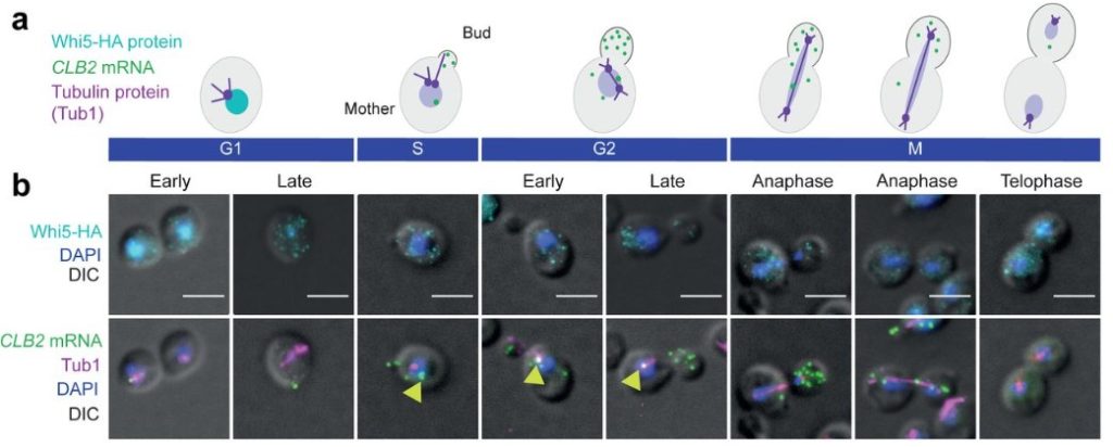
Evelina Tutucci, Anna Maekiniemi, Jacky L Snoep, Markus Seiler, Kelly van Rossum, David D. van Niekerk, Philipp Savakis, Kathi Zarnack, Robert H Singer
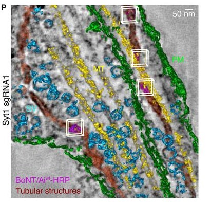
Synaptotagmin 1 mediates toxicity of botulinum neurotoxin type A
Merja Joensuu, Vanessa Lanoue, Parnayan Syed, Tristan P. Wallis, James Rae, Ailisa Blum, Rachel Gormal, Christopher Small, Shanley Sanders, Anmin Jiang, Stefan Mahrhold, Nadja Krez, Michael A. Cousin, Ruby Cooper-White, Justin J. Cooper-White, Brett A. Collins, Robert G Parton, Giuseppe Balistreri, Andreas Rummel, Frederic A Meunier
Victoria Lucia Alonso
Ana Lisica, Jonathan Fouchard, Manasi Kelkar, Tom P. J. Wyatt, Julia Duque, Anne-Betty Ndiaye, Alessandra Bonfanti, Buzz Baum, Alexandre J. Kabla, Guillaume T. Charras
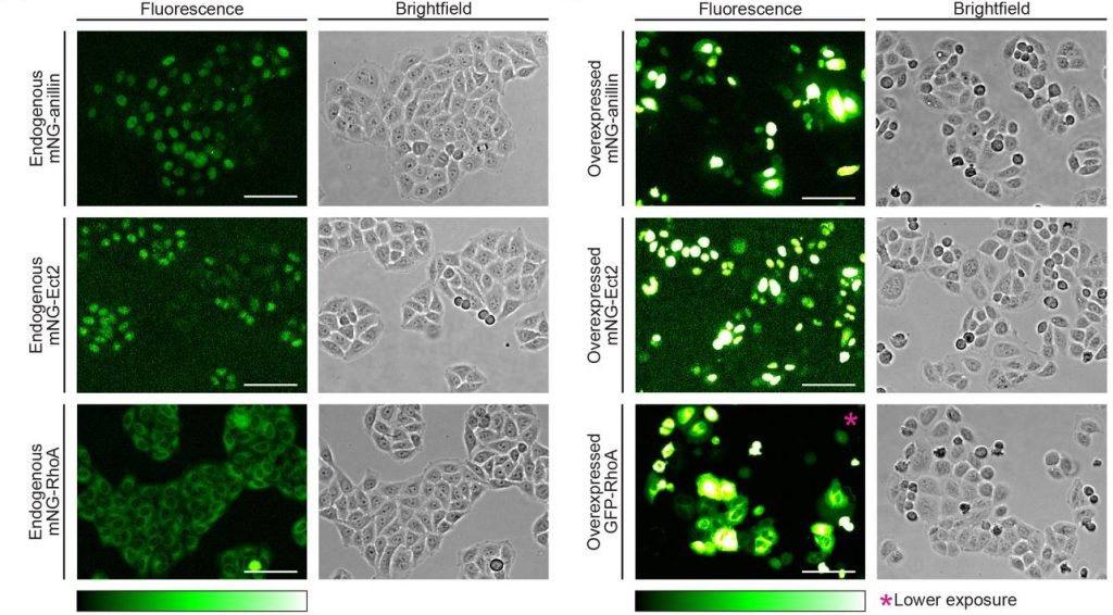
A versatile pattern-forming cortical circuit based on Rho, F-actin, Ect2, and RGA-3/4
Ani Michaud, Marcin Leda, Zachary T. Swider, Songeun Kim, Jiaye He, Jennifer Landino, Jenna R. Valley, Jan Huisken, Andrew B. Goryachev, George von Dassow, William Bement

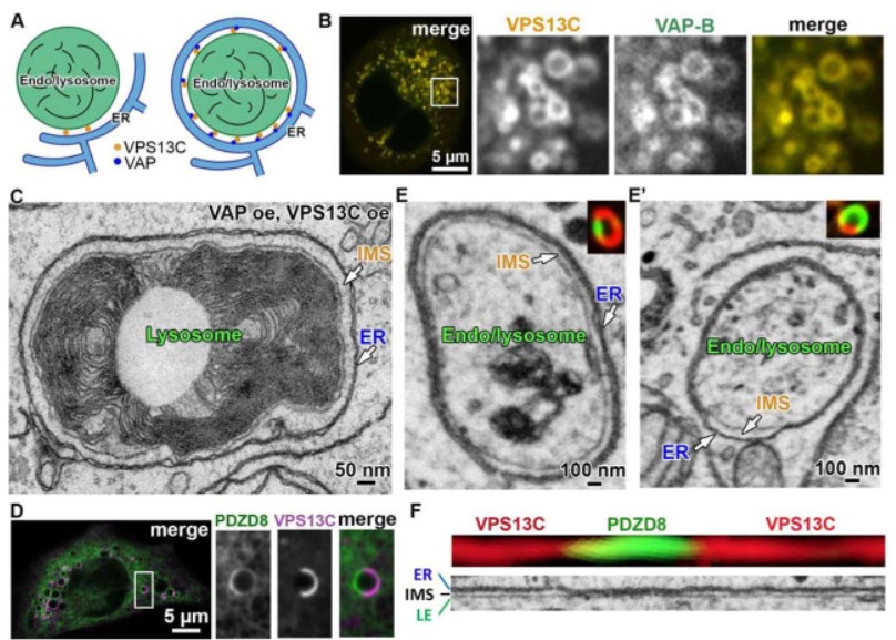
In situ architecture of the lipid transport protein VPS13C at ER-lysosomes membrane contacts
Shujun Cai, Yumei Wu, Andres Guillen-Samander, William F Hancock-Cerutti, Jun Liu, Pietro De Camilli
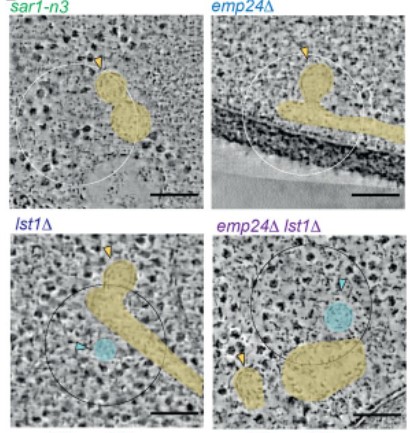
Ultrastructure of COPII vesicle formation characterised by correlative light and electron microscopy
Alejandro Melero, Jerome Boulanger, Wanda Kukulski, Elizabeth A. Miller
Imaging tools generated by CRISPR/Cas9 tagging reveal cytokinetic diversity in mammalian cells
Mathieu C. Husser, Imge Ozugergin, Tiziana Resta, Vincent J J. Martin, Alisa J. Piekny
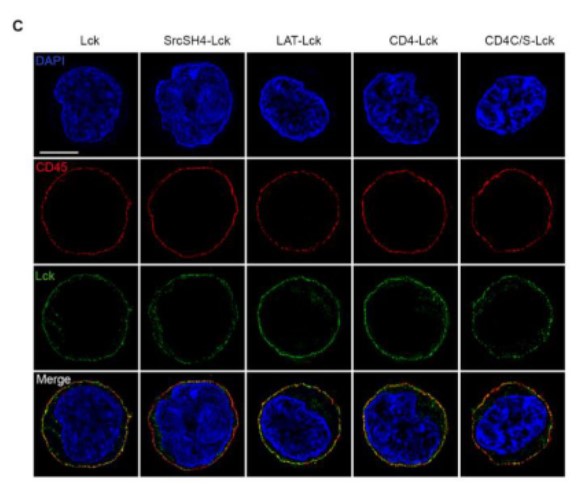
A role of Lck annular lipids in the steady upkeep of active Lck in T cells
Nicla Porciello, Deborah Cipria, Giulia Masi, Anna-Lisa Lanz, Edoardo Milanetti, Alessandro Grottesi, Duncan Howie, Steve P. Cobbold, Lothar Schermelleh, Hai-Tao He, Marco D’Abramo, Nicolas Destainville, Oreste Acuto, Konstantina Nika
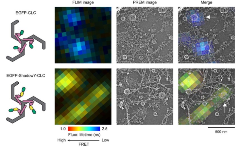
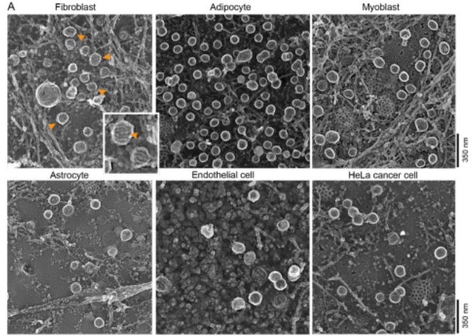
Kazuki Obashi, Kem A. Sochacki, Marie-Paule Strub, Justin W. Tarasaka
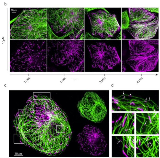
Microtubules self-repair in living cells
Morgan Gazzola, Alexandre Schaeffer, Benoit Vianay, Jérémie Gaillard, Laurent Blanchoin, Manuel Théry
Charlotte Gineste , Sonia Youhanna, Sabine U. Vorrink, Sara Henriksson, Andrés Hernández, Arthur J. Cheng, Thomas Chaillou, Andreas Buttgereit, Dominik Schneidereit, Oliver Friedrich, Kjell Hultenby, Joseph D. Bruton, Niklas Ivarsson, Linda Sandblad, Volker M. Lauschke, Håkan Westerblad
Claudia Matthaeus, Kem A. Sochacki, Andrea Dickey, Dmytro Puchkov, Volker Haucke, Martin Lehmann, Justin W. Taraska
 (No Ratings Yet)