Welcome to FocalPlane
FocalPlane is a community site for anyone who uses microscopy in their research. It is a place where you can interact with a global community of imaging scientists, engineers, chemists, and bioimage analysts.
FocalPlane is your site and once registered, you are free to share a blog post, add an image to our gallery, post a job advert or put up an event listing.
Create an account to share your story or to stay up-to-date with the latest news.
Recent posts
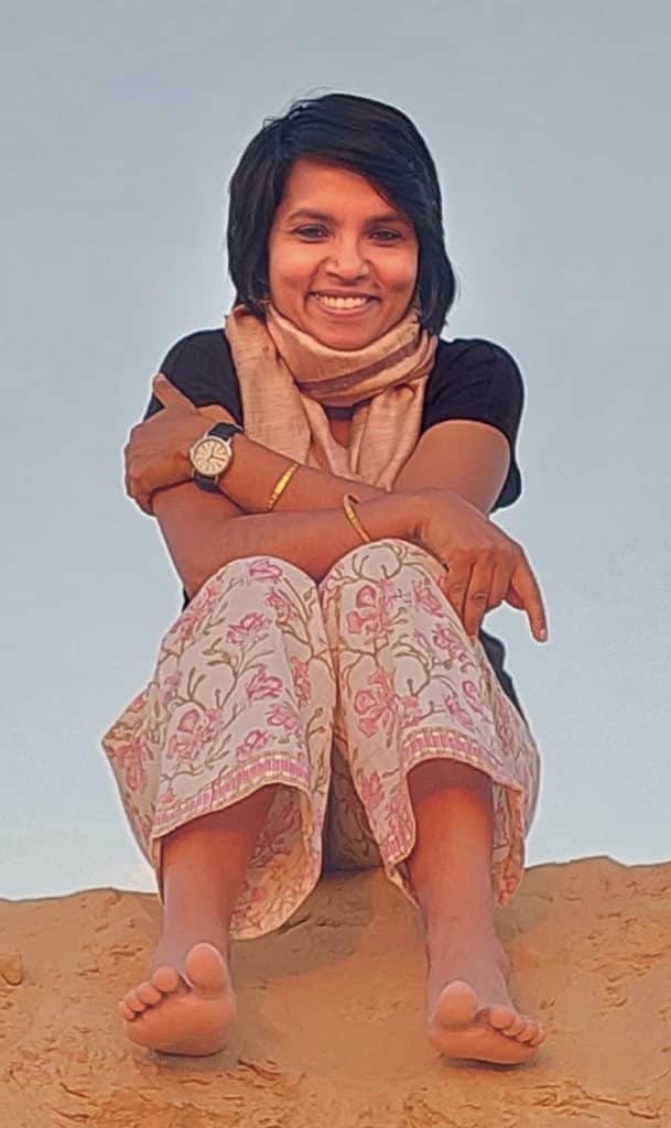
An Interview with Dr. Rashmi Priya
Posted by Subhajit Dutta, on 1 July 2025
Event carbon calculator
Posted by Teodora Andreea Rînciog, on 30 June 2025
I am excited to announce the release of our event carbon calculator, our latest resource to support the creation of sustainable events. Estimate your event’s carbon footprint in five minute or less. Our event carbon calculator provides insights into the main drivers of your meeting’s greenhouse gas emissions, offers reduction scenarios to test and providesNew strategic funding award for the UK Euro-BioImaging Node
Posted by Georgina Fletcher, on 30 June 2025
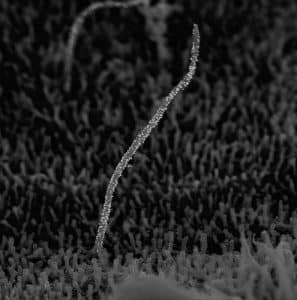
Exciting news to share with you all! The UK Euro-BioImaging Node has been awarded £1.8 million in strategic funding from UKRI-BBSRC and UKRI-MRC over five years. This transformative investment will significantly expand access to cutting-edge imaging technologies for UK researchers. What this funding will deliver: This builds on our strong foundation: 14 facilities across 7volume EM visualisation of synthetic scaffolds in bacteria
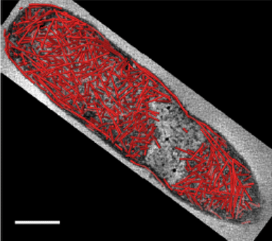
Posted by Janithri Wickramanayake, on 27 June 2025
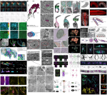
Microscopy preprints – applications in biology
Posted by FocalPlane, on 27 June 2025
Illuminating the Brain: Microscopy Techniques in Neuroscience and My Applications in the Lab
Posted by Sally Horton, on 23 June 2025
Featured image with Seth Agyei Domfeh
Posted by FocalPlane, on 20 June 2025

Most read on FocalPlane
Questions about FocalPlane?
Become a sponsor
Are you interested in becoming a sponsor for FocalPlane?
Our sister sites

The community site for
developmental biologists
Image credits
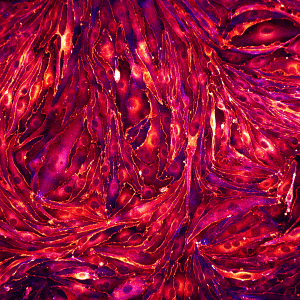
Neuromuscular junctions – Rebecca Simkins
Mitochondria and microtubules – Till Stephan
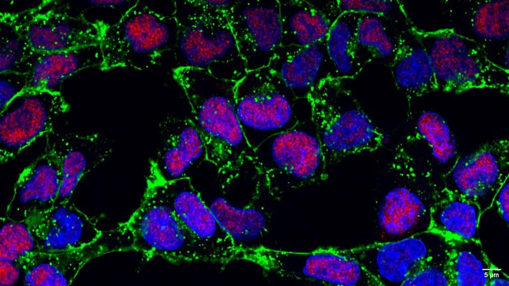
Mammary gland organoid – Oona Paavolainen
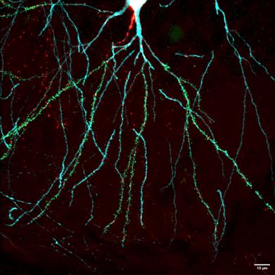
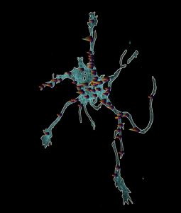
Dopaminergic neuron – Nick Gatford
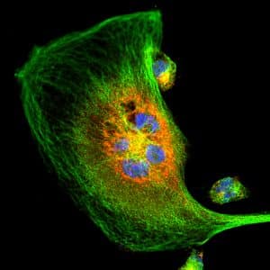
CLEM HeLa cell – Marie-Charlotte Domart, Chris Peddie
Posts by categories


Microscopy-related articles from our journals
- HCR spectral imaging: 10-plex, quantitative, high-resolution RNA and protein imaging in highly autofluorescent samples Development 2024 151: dev202307
- filoVision – using deep learning and tip markers to automate filopodia analysis J Cell Sci 2024 137: jcs261274
- Computational tools for quantifying actin filament numbers, lengths, and bundling Biology Open 2024 13: bio.060267
From the gallery

Gallery categories
- Confocal microscopyApply
- Spinning disc micros..copyApply
- Super-resolution mic..roscopyApply
- Single plane illumin..ation microscopy (SPIM / Light sheet)Apply
- Total internal refle..ction fluorescence (TIRF) microscopyApply
- Electron microscopyApply
- Multi-photon microsc..opyApply
- Label-free microscop..yApply
- DyesApply
- Genetic probesApply
- Optical manipulationApply
- Functional imagingApply